Hypertrophic cardiomyopathy (HCM) is a genetic disorder that has a variable presentation and carries a high incidence of sudden death. Its hallmark is myocardial hypertrophy that is inappropriate and often asymmetrical and that occurs in the absence of an obvious inciting hypertrophic stimulus.
Essential update: Alcohol septal ablation reduces risk in patients with hypertrophic obstructive cardiomyopathyIn a recent observational cohort study of 470 patients with obstructive HCM, echo-contrast-guided alcohol septal ablation (ASA) was associated with a low incidence of sudden cardiac death and a reduced risk profile.
The 10-year survival rate after ASA was 88%, compared with 84% in a matched control population, and the 10-year survival free of sudden cardiac death rate was 95%. ASA reduced the prevalence of syncope, abnormal blood pressure response, non-sustained ventricular tachycardia, and maximal wall thickness ≥30 mm. The procedure also reduced the proportion of patients with 2 or more risk factors for sudden cardiac death from 25% to 8%.[8]
Signs and symptomsSigns and symptoms of HCM can include the following:
Sudden cardiac death (the most devastating presenting manifestation)Dyspnea (the most common presenting symptom)Syncope and presyncopeAnginaPalpitationsOrthopnea and paroxysmal nocturnal dyspnea (early signs of congestive heart failure [CHF])CHF (relatively uncommon but sometimes seen)DizzinessPhysical findings may include the following:
Double apical impulse or triple apical impulse (less common)Normal first heart sound; second heart sound usually is normally split but is paradoxically split in some patients with severe outflow gradients; S3 gallop is common in children but signifies decompensated CHF in adults; S4 is frequently heard Jugular venous pulse revealing a prominent a waveDouble carotid arterial pulseApical precordial impulse that is displaced laterally and usually is abnormally forceful and enlargedSystolic ejection crescendo-decrescendo murmurHolosystolic murmur at the apex and axilla of mitral regurgitationDiastolic decrescendo murmur of aortic regurgitation (10% of patients)See Clinical Presentation for more detail.
DiagnosisNo specific laboratory blood tests are required in the workup. Genetic testing is not yet widely available but is becoming increasingly so.
Two-dimensional (2-D) echocardiography is diagnostic for HCM. Findings may be summarized as follows:
Abnormal systolic anterior leaflet motion of the mitral valveLeft ventricular hypertrophy (LVH)Left atrial enlargementSmall ventricular chamber sizeSeptal hypertrophy with septal-to-free wall ratio greater than 1.4:1Mitral valve prolapse and mitral regurgitationDecreased midaortic flowPartial systolic closure of the aortic valve in midsystoleOther imaging modalities that may be useful include the following:
Chest radiographyRadionuclide imagingCardiac magnetic resonance imaging: Particularly useful when echocardiography is questionable, particularly with apical hypertrophyElectrocardiographic findings may include the following:
ST-T wave abnormalities and LVH (common)Axis deviation (right or left)Conduction abnormalities (P-R prolongation, bundle-branch block)Sinus bradycardia with ectopic atrial rhythmAtrial enlargementAbnormal and prominent Q wave in the anterior precordial and lateral limb leads, short P-R interval with QRS suggestive of preexcitation, atrial fibrillation (poor prognostic sign), and a P-wave abnormality (all uncommon)The following diagnostic modalities may also be useful:
Cardiac catheterization (to determine the degree of outflow obstruction, cardiac hemodynamics, the anatomy and diastolic characteristics of the left ventricle, and the coronary anatomy) Electrophysiologic studiesSee Workup for more detail.
ManagementPharmacologic therapy for HCM may include the following:
Beta blockersCalcium channel blockersDiltiazem, amiodarone, and disopyramide (rarely)Antitussives to prevent coughingThe following caveats are warranted:
Avoid inotropic drugs if possibleAvoid nitrates and sympathomimetic amines, except in concomitant coronary artery diseaseAvoid digitalisUse diuretics with cautionSurgical and catheter-based therapeutic options include the following:
Left ventricular myomectomyMitral valve replacementPermanent pacemaker implantationCatheter septal ablationPlacement of an implantable cardioverter defibrillatorSee Treatment and Medication for more detail.
Image library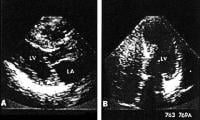 Hypertrophic cardiomyopathy. NextBackground
Hypertrophic cardiomyopathy (HCM) is a genetic disorder that is typically inherited in an autosomal dominant fashion with variable penetrance and variable expressivity. The disease has complex symptomatology and potentially devastating consequences for patients and their families. (See Etiology and Prognosis.)[1]
The disorder has a variable presentation and carries a high incidence of sudden death. In fact, HCM is the leading cause of sudden cardiac death in preadolescent and adolescent children. The hallmark of the disorder is myocardial hypertrophy that is inappropriate, often asymmetrical, and occurs in the absence of an obvious inciting hypertrophy stimulus. This hypertrophy can occur in any region of the left ventricle but frequently involves the interventricular septum, which results in an obstruction of flow through the left ventricular (LV) outflow tract. (See Prognosis, History, Physical Examination, and Workup.)
Decades ago, HCM was written about and known as idiopathic hypertrophic subaortic stenosis (IHSS) or asymmetrical septal hypertrophy (ASH). These terms were replaced by hypertrophic cardiomyopathy, because the segmental hypertrophy can occur in any segment of the ventricle, not just the septum. Furthermore, this entity can present without subaortic obstruction to flow yet still carry the same ominous risk of arrhythmogenic sudden death and many of its clinical symptoms.
HCM can be separated into obstructive and nonobstructive types. Obstructive HCM is due to midsystolic obstruction of flow through the LV outflow tract as a result of a Bernoulli effect–induced systolic anterior mitral valve movement toward the septum.
The significance of this obstruction, however, is highly controversial. Some investigators and experts believe the obstruction has less to do with the overall hemodynamic and pathophysiologic manifestations of this entity than it does with the inappropriate segmental hypertrophy, which, with its increased myocardial oxygen consumption and substrate for fatal ventricular arrhythmias, has much more significance in the overall clinical picture of this entity and in the treatment and prognosis of HCM.
HCM is a familial disease.[2] There are defects in several of the genes encoding for the sarcomeric proteins, such as myosin heavy chain, actin, tropomyosin, and titin.[3, 4] Multiple mutations have been identified, with genotype-specific risks for mortality and degree of hypertrophy. Interestingly, the genetic basis of ventricular hypertrophy does not directly correlate with prognostic risk stratification. (See Etiology.)
Patients with some mutations, such as specific tropomyosin substitutions, have only a mild degree of ventricular hypertrophy, with little or no LV outflow tract obstruction, but they still carry a disproportionately high risk for sudden death.[5]
Many patients, particularly children, with HCM may not be symptomatic. Careful evaluation of a heart murmur may reveal the condition. (See Physical Examination and Workup.)
2011 American College of Cardiology Foundation (ACCF)/AHA Guideline for the Diagnosis and Treatment of Hypertrophic Cardiomyopathy is now available.[6, 7]
ComplicationsComplications of HCM may include the following:
Congestive heart failureVentricular and supraventricular arrhythmiasInfective mitral endocarditisAtrial fibrillation with mural thrombus formationSudden deathPreviousNextPathophysiologySince the initial descriptions of hypertrophic cardiomyopathy (HCM), the feature that has attracted the greatest attention is the dynamic pressure gradient across the LV outflow tract. The pressure gradient appears to be related to further narrowing of an already small outflow tract (already narrowed by the marked asymmetrical septal hypertrophy and possibly by an abnormal location of the mitral valve) by the systolic anterior motion of the mitral valve against the hypertrophied septum.
Three explanations for the systolic anterior motion of the mitral valve have been offered, as follows: (1) the mitral valve is pulled against the septum by contraction of the papillary muscles, which occurs because of the valve's abnormal location and septal hypertrophy altering the orientation of the papillary muscles; (2) the mitral valve is pushed against the septum because of its abnormal position in the outflow tract; (3) the mitral valve is drawn toward the septum because of the lower pressure that occurs as blood is ejected at high velocity through a narrowed outflow tract (Venturi effect).
Most patients with HCM have abnormal diastolic function (whether or not a pressure gradient is present), which impairs ventricular filling and increases filling pressure, despite a normal or small ventricular cavity. These patients have abnormal calcium kinetics and subendocardial ischemia, which are related to the profound hypertrophy and myopathic process.
PreviousNextEtiologyAbnormal calcium kineticsData link abnormal myocardial calcium kinetics to the cause of the inappropriate myocardial hypertrophy and specific features of HCM, particularly in patients with diastolic functional abnormalities. Abnormal myocardial calcium kinetics and abnormal calcium fluxes from an increase in the number of calcium channels result in an increase in intracellular calcium concentration, which, in turn, may produce hypertrophy and cellular disarray.
Genetic causesFamilial HCM occurs as an autosomal dominant Mendelian-inherited disease in approximately 50% of cases. Some, if not all, of the sporadic forms of the disease may be caused by spontaneous mutations.
At least 6 different genes on at least 4 chromosomes are associated with HCM, with more than 50 different mutations discovered thus far. Familial HCM is a genetically heterogenous disease in that it can be caused by genetic defects at more than 1 locus.
In 1989, Seidman and collaborators first reported the genetic basis for HCM. They reported the existence of a disease gene located on the long arm of chromosome 14. Subsequently, they found this to be the gene encoding for beta cardiac myosin heavy chain.
Wide variation exists in the phenotypic expression of a given mutation of a given gene, with variability in clinical symptoms and the degree of hypertrophy expressed. Phenotypic variability is related to the differences in genotype, with specific mutations associated with particular symptoms, the degree of hypertrophy, and the prognosis.[9]
Other possible causesOther possible causes of HCM include the following:
Abnormal sympathetic stimulation - Heightened responsiveness of the heart to the excessive production of catecholamines or the reduced neuronal uptake of norepinephrine might cause HCM Abnormally thickened intramural coronary arteries - These do not dilate normally, which leads to myocardial ischemia; this progresses to myocardial fibrosis and abnormal compensatory hypertrophy Subendocardial ischemia - This is related to abnormalities of the cardiac microcirculation that deplete the energy stores essential for the sequestration of calcium during diastole; subendocardial ischemia results in persistent interaction of the contractile elements during diastole and increased diastolic stiffness Cardiac structural abnormalities - These include a catenoid configuration of the septum, which results in myocardial cell hypertrophy and disarray PreviousNextEpidemiologyHypertrophic cardiomyopathy (HCM) is reported in 0.5% of the outpatient population referred for echocardiography.[10] The overall prevalence of HCM is low and has been estimated to occur in 0.05-0.2% of the population.[11] Morphologic evidence of disease is found by echocardiography in approximately 25% of first-degree relatives of patients with HCM. Genetic testing still is in the early stages of research development but can be used to identify asymptomatic family members with the same mutation as the proband (index case).
Sex-related demographicsHCM is slightly more common in males than in females. However, the genetic inheritance pattern is autosomal dominant, without sex predilection. Modifying genetic, hormonal, and environmental factors may lead to a higher likelihood of identification in males, increased symptomatology, or higher degrees of LV outflow obstruction, with more prominent findings upon physical examination.
HCM usually presents at a younger age in females. Females tend to be more symptomatic and are more likely to be disabled by their symptoms than males.
Age-related demographicsIn general, HCM has a bimodal peak of occurrence. The most common presentation is in the third decade of life, but it may present in persons of any age, from newborns to elderly individuals.
In children, inherited cases are found in an age range from newborn (ie, stillborn babies) to adult. The peak incidence is in these cases is in the second decade of life.
In adults, the peak incidence is in the third decade of life, with the vast majority of cases occurring in the age range between the third and sixth decades of life.
PreviousNextPrognosisReported annual mortality rates in patients with hypertrophic cardiomyopathy (HCM) have ranged from less than 1% to 3-6%, and studies suggest that they have significantly improved over the past 40 years.[12]
A 2006 study reported that published sudden death rates over the previous 10 years were lower than were previously published figures (median 1.0% (range 0.1–1.7) v 2.0% (0–3.5)). Nevertheless, HCM still carries a high risk for mortality and morbidity.[13]
One series of 46 patients with midventricular obstruction was found to have an increased risk of apical aneurysm formation, symptoms, and HCM-related death compared with those who did not have midventricular obstruction; the increased risk of symptoms and death was similar to that seen in patients with LV outflow obstruction.[14]
Most patients with HCM are asymptomatic. Unfortunately, the first clinical manifestation of the disease in such individuals may be sudden death, likely from ventricular tachycardia or fibrillation. Younger patients, particularly children, have a much higher mortality rate. Children have a much greater degree of ventricular hypertrophy and are much more symptomatic early on in the disease course, most likely because more malignant genotypes are present earlier in life.
The more benign mutations do not elicit a clinical or echocardiographic phenotype or symptoms in the pediatric population. Death often is sudden, unexpected, and typically is associated with sports or vigorous exertion. Early diagnosis is of prime importance in order to prescribe an appropriate level of safe activity.[9, 15, 16]
Screening of first-degree relatives is useful to identify additional affected family members prior to the onset of significant symptoms or sudden death.
Patients can have a myriad of arrhythmias, including atrial fibrillation, atrial flutter, ventricular ectopy, ventricular tachycardia, and ventricular fibrillation. These patients are among the highest-risk group for ventricular fibrillation and pose difficult therapeutic decisions for risk reduction.
Patients have a high likelihood of recurrent heart failure resulting from mitral regurgitation and profound diastolic dysfunction. HCM is a progressive condition that worsens over time, as does the gradient across the LV outflow tract if left untreated. Systolic function usually is well preserved until the late stages of the disease. Angina is rare in children but common in adults. Syncope and presyncope are common and may identify individuals at high risk for sudden death.
PreviousNextPatient EducationFamily members should learn cardiopulmonary resuscitation. In addition, refer the patient and family for psychosocial counseling. Refer children of patients with hypertrophic cardiomyopathy (HCM), especially those in the pediatric age range, for urgent echocardiography and genetic testing if an echocardiogram does not yet reveal overt disease.
Impose activity restrictions that include total abstinence from highly competitive athletic activities and very strenuous physical exertion, such as lifting heavy objects, lifting weights, and shoveling snow.
For patient education information, see the Heart Health Center, as well as Palpitations.
PreviousProceed to Clinical Presentation , Hypertrophic Cardiomyopathy


0 comments:
Post a Comment