Cardiac tamponade is a time sensitive, life-threatening condition that requires prompt diagnosis and management. Historically, the diagnosis of cardiac tamponade has been based on clinical findings. Claude Beck, a cardiovascular surgeon, described 2 triads of clinical findings that he found associated with acute and chronic cardiac tamponade. The first of these triads consisted of hypotension, an increased venous pressure, and a quiet heart. It has come to be recognized as Beck's triad, a collection of findings most commonly produced by acute intrapericardial hemorrhage. Subsequent studies have shown that these classic findings are observed in only a minority of patients with cardiac tamponade.[1]
The detection of pericardial fluid has been facilitated by the development and continued improvement of echocardiography. Cardiac ultrasound is now accepted as the criterion standard imaging modality for the assessment of pericardial effusions and the dynamic findings consistent with cardiac tamponade. With echocardiography, the location of the effusion can be identified, the size can be estimated (small, medium, or large), and the hemodynamic effects can be examined by assessing for abnormal septal motion, right atrial or right ventricular inversion, and decreased respiratory variation of the diameter of the inferior vena cava.[2]
Subxiphoid views of pericardial effusion are shown below.
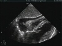 Subxiphoid view of the heart demonstrating a moderate sized pericardial effusion.
 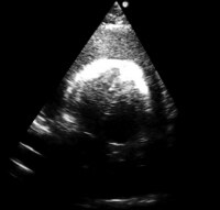 Subxiphoid view of the heart demonstrating a large pericardial effusion.
 The video below depicts subxiphoid cardiac tamponade.
Cine loop depicting subxiphoid cardiac tamponade. Video courtesy of Meghan Kelly Herbst, MD. Also courtesy of Yale School of Medicine, Emergency Medicine.Pericardiocentesis is the aspiration of fluid from the pericardial space that surrounds the heart. This article describes the landmark or “blind†syringe and needle technique used as a lifesaving measure for the prompt management of cardiac tamponade.
Historical backgroundPercutaneous pericardiocentesis was introduced during the 19th century. Frank Schuh first described this procedure in 1840. By the 20th century, percutaneous pericardiocentesis became a preferred technique for the treatment of patients with pericardial effusion or for diagnostic purposes.
Before the advent of 2-dimensional echocardiography, the procedure used a blind-subxiphoid approach. Serious complications were not uncommon (eg, injury to liver, myocardium, coronary arteries, lungs). Because 2-dimensional echocardiography permits direct visualization of cardiac structures and adjacent vital organs, the procedure now is performed with minimal risk. Since 1979, echo-guided pericardiocentesis has been the preferred initial procedure for the diagnosis and treatment of most pericardial effusions. The technique has been modified and refined in the past 22 years. Percutaneous pericardiocentesis now is the procedure of choice for the safe removal of pericardial fluid. Whenever possible, this procedure should be performed by a surgeon, an interventional cardiologist or a cardiologist trained in invasive techniques.
There is no significant difference in overall mortality between open surgical drainage and percutaneous pericardiocentesis for symptomatic pericardial effusions. There may be more procedural complications following surgical drainage of a pericardial effusion, and the need for repeat procedures may be greater if the effusion is drained using pericardiocentesis.[3]
Next, Pericardiocentesis


0 comments:
Post a Comment