Cardiac tamponade is a clinical syndrome caused by the accumulation of fluid in the pericardial space, resulting in reduced ventricular filling and subsequent hemodynamic compromise. The condition is a medical emergency, the complications of which include pulmonary edema, shock, and death. (See Pathophysiology, Etiology, and Prognosis.)
The overall mortality risk depends on the speed of diagnosis, the treatment provided, and the underlying cause of the tamponade. Untreated, the condition is rapidly and universally fatal (see the image below). (See Presentation, Workup, Treatment, and Medication.)
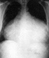 This anteroposterior-view chest radiograph shows a massive, bottle-shaped heart and conspicuous absence of pulmonary vascular congestion. Reproduced with permission from Chest, 1996: 109:825. NextPathophysiology
The pericardium, which is the membrane surrounding the heart, is composed of 2 layers. The thicker parietal pericardium is the outer fibrous layer; the thinner visceral pericardium is the inner serous layer. The pericardial space normally contains 20-50mL of fluid.
Reddy et al describe 3 phases of hemodynamic changes in tamponade.[1]
Phase I - The accumulation of pericardial fluid causes increased stiffness of the ventricle, requiring a higher filling pressure; during this phase, the left and right ventricular filling pressures are higher than the intrapericardial pressure Phase II - With further fluid accumulation, the pericardial pressure increases above the ventricular filling pressure, resulting in reduced cardiac output (see the Cardiac Output calculator) Phase III - A further decrease in cardiac output occurs, which is due to the equilibration of pericardial and left ventricular (LV) filling pressuresPericardial effusions, which cause cardiac tamponade, can be serous, serosanguineous, hemorrhagic, or chylous.
The underlying process for the development of tamponade is a marked reduction in diastolic filling, which results when transmural distending pressures become insufficient to overcome increased intrapericardial pressures. Tachycardia is the initial cardiac response to these changes to maintain the cardiac output.
Systemic venous return is also altered during tamponade. Because the heart is compressed throughout the cardiac cycle due to the increased intrapericardial pressure, systemic venous return is impaired and right atrial and right ventricular collapse occurs. Because the pulmonary vascular bed is a vast and compliant circuit, blood preferentially accumulates in the venous circulation, at the expense of LV filling. This results in reduced cardiac output and venous return.
The amount of pericardial fluid needed to impair diastolic filling of the heart depends on the rate of fluid accumulation and the compliance of the pericardium. Rapid accumulation of as little as 150mL of fluid can result in a marked increase in pericardial pressure and can severely impede cardiac output,[2] whereas 1000 mL of fluid may accumulate over a longer period without any significant effect on diastolic filling of the heart. This is due to adaptive stretching of the pericardium over time. A more compliant pericardium can allow considerable fluid accumulation over a longer period without hemodynamic insult.
PreviousNextEtiologyFor all patients, malignant diseases are the most common cause of pericardial tamponade. Among etiologies for tamponade, Merce et al reported the following incidence rates:
Malignant diseases - 30-60% of casesUremia - 10-15% of casesIdiopathic pericarditis - 5-15%Infectious diseases - 5-10%Anticoagulation - 5-10%Connective tissue diseases - 2-6%Dressler or postpericardiotomy syndrome - 1-2%Tamponade can occur as a result of any type of pericarditis. Pericarditis can result from the following[3] :
Human immunodeficiency virus (HIV) infectionInfection - Viral, bacterial (tuberculosis), fungalDrugs - Hydralazine, procainamide, isoniazid, minoxidilPostcoronary intervention - Ie, coronary dissection and perforationAcupuncture[4] Postcardiac percutaneous procedures - Including mitral valvuloplasty, atrial septal defect (ASD) closure, left atrial appendage occlusion Trauma to the chestCardiovascular surgery - Postoperative pericarditis[5] Postmyocardial infarction - Free wall ventricular rupture, Dressler syndromeConnective tissue diseases - Systemic lupus erythematosus, rheumatoid arthritis, dermatomyositisRadiation therapy to the chestIatrogenic[6] - After sternal biopsy, transvenous pacemaker lead implantation, pericardiocentesis, or central line insertion UremiaAnticoagulation treatmentIdiopathic pericarditisComplication of surgery at the esophagogastric junction - Eg, antireflux surgeryPneumopericardium - Due to mechanical ventilation or gastropericardial fistulaHypothyroidismStill diseaseDuchenne muscular dystrophyType A aortic dissectionPreviousNextEpidemiologyOccurrence in the United StatesThe incidence of cardiac tamponade is 2 cases per 10,000 population in the United States. Approximately 2% of penetrating injuries are reported to result in cardiac tamponade.
Sex- and age-related demographicsIn children, cardiac tamponade is more common in boys than in girls, with a male-to-female ratio of 7:3. In adults, cardiac tamponade appears to be slightly more common in men than in women. A male-to-female ratio of 1.25:1 was observed at the author's referral center, based on the International Classification of Diseases (ICD) code 423.9. However, a male-to-female ratio of 1.7:1 was observed at another level 1 trauma center.
Cardiac tamponade related to trauma or HIV is more common in young adults, whereas tamponade due to malignancy and/or renal failure occurs more frequently in elderly individuals.
PreviousNextPrognosisCardiac tamponade is a medical emergency. The prognosis depends on prompt recognition and management of the condition and the underlying cause of the tamponade. Untreated, cardiac tamponade is rapidly and universally fatal.
In addition to treatment for the tamponade, all patients should also receive treatment for the condition’s underlying cause in order to prevent recurrence.
In a study of patients with cardiac tamponade, Cornily et al reported a 1-year mortality rate of 76.5% in patients whose tamponade was caused by malignant disease, compared with 13.3% in patients with no malignant disease. The investigators also noted a median survival of 150 days in patients with malignant disease.[7]
PreviousProceed to Clinical Presentation , Cardiac Tamponade


0 comments:
Post a Comment