Peripartum cardiomyopathy (PPCM) has a number of definitions, but the authors prefer to use the one put forth by the Heart Failure Association of the European Society of Cardiology Working Group on PPCM 2010.[1]
By this definition, PPCM is an idiopathic cardiomyopathy that presents with heart failure secondary to left ventricular systolic dysfunction toward the end of pregnancy or in the months after delivery, in the absence of any other cause of heart failure. PPCM is a diagnosis of exclusion. Although the left ventricle may not be dilated, the ejection fraction is nearly always reduced below 45%.[1]
In contrast to other definitions, the Heart Failure Association’s definition specifically excludes women who develop PPCM early in their pregnancy and explicitly notes that not all cases of PPCM present with left ventricular dilation.[2] Thus, it helps avoid misdiagnosis of other conditions that present with pulmonary edema in pregnancy, such as diastolic dysfunction from preeclampsia and other disorders (see Diagnosis).
PPCM is more common in multiparous women. It has been reported more often in twin gestations and in women with preeclampsia, but both of these conditions are associated with a lower serum oncotic pressure that can predispose to noncardiogenic pulmonary edema in the setting of other stressors.
The severity of symptoms in patients with PPCM can be classified by the New York Heart Association system as follows:
Class I - Disease with no symptomsClass II - Mild symptoms/effect on function or symptoms only with extreme exertionClass III - Symptoms with minimal exertionClass IV - Symptoms at restNextPathophysiology and EtiologyThe exact cause of peripartum cardiomyopathy (PPCM) is unknown, but the usual causes of systolic dysfunction and pulmonary edema should be excluded. Many nutritional disorders have been suggested as causes, but other than salt overload, none has been validated by epidemiologic studies.
An increased prevalence of myocarditis has been found in case series and in a small case-control study. Abnormal myocardial biopsy findings were associated with a worse long-term prognosis for recovery. More recent data have found a similar incidence of myocarditis in women with PPCM, compared to those with the idiopathic type. However, a study that found myocarditis in 62% of 44 women with PPCM found that the finding did not correlate with survival.
Lower levels of selenium have been found in patients with PPCM. Autoantibodies against myocardial proteins have been identified in patients with PPCM but not in those with idiopathic cardiomyopathy.[3]
Case reports and anecdotal experience have documented ejection fractions as low as 10-15% in patients with severe preeclampsia, with subsequent normalization of echocardiograms within 3-6 months. Preeclampsia has been listed as a risk factor, but it may be the cause in some cases. Noncardiogenic pulmonary edema has many causes, all of which must be considered.[4]
A study in 2005 found that 8 of 26 patients had parvovirus B19, human herpes virus 6, Epstein-Barr virus, and human cytomegalovirus detected after molecular analysis of myocardial biopsy specimens.[5]
Other findings that are associated with PPCM but are not clearly causal include increased levels of inflammation and oxidative stress markers, increased levels of cathepsin D, and oxidized low-density lipoprotein.
Two studies have suggested that a subset of cases of PPCM result from a genetic cause.[6, 7] Case reports of women from the same family who developed PPCM suggest a possible familial/genetic risk, but it seems that some of these women may have familial dilated cardiomyopathy that is unmasked by the normal physiologic changes of pregnancy.[6]
Some have hypothesized that microchimerism, or fetal cells present in the maternal system that elicit an inflammatory response, could be a potential contributing factor to the development of PPCM.
PreviousNextEpidemiologyUnited States statisticsReports estimating the incidence of peripartum cardiomyopathy (PPCM) in the United States vary widely, ranging from 1 case per 15,000 live births to 1 case per 4000 live births to 1 case per 1300 live births. Approximately 75% of cases are diagnosed within the first month post partum, and 45% present in the first week. When PPCM is suspected, one must establish the diagnosis rapidly.[8]
International statisticsThe prevalence is reported to be 1 case per 6000 live births in Japan, 1 case per 1000 live births in South Africa, and 1 case per 350-400 live births in Haiti. A high prevalence in Nigeria is caused by the tradition of ingesting kanwa (dried lake salt) while lying on heated mud beds twice a day for 40 days post partum. The high salt intake leads to volume overload.
Age-, sex-, and race-related demographicsPPCM is unique to pregnant women of all reproductive ages. Initially thought to be more common in women older than 30 years, PPCM has since been reported across a wide range of age groups. The past bias toward older women may be related to the fact that this group has a higher prevalence of undiagnosed conditions, such as thyrotoxicosis, mitral stenosis, or hypertension, which, in combination with some complication of pregnancy and the physiologic alterations of pregnancy, leads to pulmonary edema.
PPCM has been reported in white, Chinese, Korean, and Japanese women. Case series indicate that many cases occur in African American women from the southern United States. A case control study in the United States found that, when compared to non-African Americans, African American women had a 15.7-fold higher relative risk of PPCM.[9]
PreviousNextClinical PresentationPatient historyMany presenting complaints observed in patients with cardiac disease occur during a normal pregnancy. Dyspnea, dizziness, orthopnea, and decreased exercise capacity often are normal symptoms in pregnant women. Mild dyspnea upon exertion is particularly common in a normal pregnancy. The classic dyspnea of pregnancy is often described as the woman feeling as if she is unable to get enough air in, to get a good deep breath, or both, and it is thought to be due to the progesterone-mediated hyperventilation.
Early, rapid diagnosis of peripartum cardiomyopathy (PPCM) is not the norm. It took 7 or more days to establish the diagnosis in 48% of women, and half of those had major adverse events before the diagnosis was made.[10] Often, patients do not show any indication of the syndrome until after delivery. If a patient makes it through labor—essentially nature’s stress test—without symptoms, the onsite clinician might not consider PPCM as the first cause when a woman decompensates.
Many PPCM patients present with heart failure or a major adverse event (eg, stroke or respiratory failure) without any previous signs or symptoms to alert the clinician that a cardiomyopathy was going to develop; 19% of patients may present with the syndrome before the last gestational month.[8] Symptoms are the same as in patients with systolic dysfunction who are not pregnant. New or rapid onset of the following symptoms requires prompt evaluation:
CoughOrthopneaParoxysmal nocturnal dyspneaFatiguePalpitationsHemoptysisChest painAbdominal painPhysical examinationIn a normal pregnancy, as a result of the increase in endogenous progestins, respiratory tidal volume is increased and patients have a tendency to hyperventilate. However, the rate of respiration should be normal. Normal pregnancy is characterized by an exaggerated x and y descent of the jugular venous waveform, but the jugular venous pressure should be normal.
Cardiac auscultation reveals a systolic ejection murmur at the lower left sternal edge, over the pulmonary area, or both in 96% of women.[11] This pulmonic arterial flow murmur tends to become quieter during inspiration. Diastolic murmurs warrant further evaluation.
The first heart sound (S1) may be exaggerated, and the second heart sound (S2) split may be more prominent due to increased right-sided flow. Whereas a third heart sound (S3) has been described as a normal finding in pregnancy, the authors have not found that to be the case in busy clinical practices at women’s hospitals that see approximately 14,000 deliveries a year.
Peripheral edema occurs in approximately one third of healthy gravid women. However, be alert to sudden changes in swelling late in pregnancy, which can be abnormal and should be investigated.
In a patient with PPCM, signs of heart failure are the same as in patients with systolic dysfunction who are not pregnant. Tachycardia and decreased pulse oximetry (should be ≥ 97% at sea level) are present. Blood pressure may be normal. Elevated blood pressures (systolic >140 mm Hg and/or diastolic >90 mm Hg) and hyperreflexia with clonus suggest preeclampsia.
Physical findings of PPCM include elevated jugular venous pressure, cardiomegaly, third heart sound, loud pulmonic component of the second heart sound, mitral or tricuspid regurgitation, pulmonary rales, worsening of peripheral edema, ascites, arrhythmias, embolic phenomenon, and hepatomegaly.
Maternal and fetal complicationsMaternal complications may include the following:
HypoxiaThromboembolism - Small series have reported the incidence to be as high as 50%, but these results have obvious selection bias Progressive cardiac failureArrhythmiasMisinterpretation of hemodynamic data obtained from right-heart catheterization as a result of failure to consider the normal physiologic alterations of pregnancy (see Invasive Hemodynamic Monitoring) Inadequate treatment or testing because of exaggerated concern about the effect on the fetusMisdiagnosis of preeclampsia - Patients with preeclampsia experience depletion of intravascular volume and should receive low doses of diuretics only when they have pulmonary edemaFetal complications may include the following:
Distress due to maternal hypoxiaDistress due to placental hypoperfusion as a result of poor cardiac output, maternal hypovolemia due to excessive diuresis, or hypotension from aggressive afterload reduction PreviousNextDiagnosisThe differential diagnosis includes the following:
Aortic StenosisCardiomyopathy, AlcoholicCardiomyopathy, CocaineCardiomyopathy, DilatedCardiomyopathy, HypertrophicCardiomyopathy, RestrictiveCardiovascular Disease and PregnancyCoronary Artery AtherosclerosisHypertensionHypertension and PregnancyHypertension, MalignantMitral StenosisNoncardiogenic pulmonary edema during pregnancy: Pregnancy is a state of low oncotic pressure reflected in decreased serum albumin (expected values, ~3.2 mg/dL); consequently, when other stressors are present, pulmonary edema can occur with normal cardiac filling pressures; the most common triggers include pyelonephritis and other infections, corticosteroids, and tocolytics such as beta agonists and magnesium sulfate Preeclampsia (toxemia of pregnancy)Pulmonary Disease and PregnancyPulmonary Edema, CardiogenicPulmonary Edema, NeurogenicOther problems to be considered include the following:
Arrhythmogenic right ventricular dysplasiaCardiomyopathy, diabetic heart diseaseInfectious, toxic, or metabolic disordersPreviousNextLaboratory StudiesCreatinine phosphokinase (CPK) levels can be elevated after normal delivery due to release from the uterus and may be elevated after cesarean section due to release from the uterus and/or skeletal muscle. An elevated CPK level is not diagnostic of peripartum cardiomyopathy (PPCM), because it can be elevated for many other reasons, including normal delivery, skeletal muscle disorders, and viral myocarditis.
The CPK from the placenta routinely has a CPK-MB fraction of 6% or more.[12] Therefore, without an obvious clinical presentation and electrocardiographic (ECG) findings to suggest myocardial infarction, the use of this test in the puerperium is very limited.
Troponin-I elevations are more likely to indicate true myocardial disease, whether it is inflammatory or due to infarction. They are certainly useful in diagnosing acute myocardial infarction.
One study found that a cardiac troponin T level greater than 0.04 ng/mL, measured within 2 weeks of diagnosis, was 60% sensitive at identifying women more likely to have persistent ventricular dysfunction at 6 months after the diagnosis. Given the poor sensitivity, the clinical use of this test is not entirely clear; these women should be placed on maximal medical therapy and undergo serial echocardiographic assessments regardless of the troponin result.[13]
Preeclampsia should be excluded on the basis of the history, the physical examination, and blood work. New headaches, visual disturbances, right-side abdominal pain, and new swelling of the hands or face may be present. Retinal vasospasm, a fourth heart sound (S4) heard on cardiac auscultation, hyperreflexia/clonus, right upper quadrant tenderness, and face or hand edema may be present. Abnormalities found with preeclampsia include the following:
Serum creatinine level greater than 0.8 mg/dLHemoglobin level greater than 13 g/dl (due to leaky capillaries and hemoconcentration)Elevated liver enzymesThrombocytopeniaUrine dipstick test results indicating more than “1+†proteinDecreased 24-hour urine creatinine clearance (normally 150% above the nonpregnant level, or approximately 150 mL/min)More than 300 mg of proteinuria evident on a 24-hour collectionOn urinalysis, trace or 1+ proteinuria can be normal. Proteinuria 2+ or higher suggests preeclampsia. Exclude infection; urine culture is helpful in this regard.
Measure pulse oximetry. Determine thyroid-stimulating hormone levels. Keep the potassium level above 4 mEq/L and the magnesium level above 2 mEq/L.
Serologic testing may help identify known causes of cardiomyopathy, including infections (eg, viral, rickettsial, HIV, syphilis, Chagas disease, diphtheria toxin). Exclude toxic etiologies such as ethanol and cocaine. When indicated, exclude systemic disorders such as collagen vascular diseases, sarcoidosis, thyrotoxicosis, pheochromocytoma, and acromegaly.
ElectrocardiographyECGs evaluate for conduction abnormalities. Results may be normal, show sinus tachycardia, or, rarely, atrial fibrillation if the cardiomyopathy is severe. Other nonspecific findings include low voltage, left ventricular hypertrophy, and nonspecific ST-segment and T-wave abnormalities.
PreviousNextEchocardiographyConsider calling a cardiologist for an immediate echocardiogram. Echocardiography should be performed in all women in whom the diagnosis of PPCM is considered in order to assess ventricular function, valve structure, chamber size, and wall motion. Cardiac chambers enlarge slightly during pregnancy, usually within normal limits. Normal function suggests a lung process or noncardiogenic pulmonary edema, and diastolic dysfunction can be observed in patients with severe preeclampsia.
PreviousNextChest RadiographyRapid diagnosis must be established. When evaluating new onset dyspnea, tachycardia, or hypoxia, immediately obtain a chest radiograph to detect pulmonary edema. This should be performed with abdominal shielding to evaluate the etiology of hypoxia and exclude pneumonia.
Fetal radiation exposure with 2 maternal chest radiographs with abdominal shielding is about 0.00007 rads. The accepted limit of fetal radiation exposure during pregnancy is 5 rads. To reassure patients about the safety of a single study, note that you are obtaining 1 out of more than 70,000 maternal chest radiographs that are theoretically permissible.
Patchy infiltrates in the lower lung fields, with vascular redistribution/cephalization, cardiomegaly, and pleural effusions, indicate congestive heart failure. Remember that noncardiogenic pulmonary edema may occur when a pregnant woman has a concurrent infection. In this setting, the cardiac pressures may be normal and cephalization of vessels may not be present.
Bilateral lower lobe infiltrates without vascular redistribution suggest either an atypical pneumonia or noncardiogenic pulmonary edema (see the image below) resulting from the low oncotic state of pregnancy combined with various stressors.
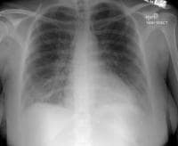 Noncardiogenic pulmonary edema in patient with preeclampsia, due to capillary leak that can be primary component of preeclampsia. Radiograph reveals diffuse increase in lung markings without cephalization or vascular redistribution seen in patients with pulmonary edema from systolic dysfunction. Patient had rapid clinical improvement after only 10 mg of intravenous furosemide. PreviousNextStress Testing
Exclude coronary artery disease when the patient presents with symptoms suggestive of cardiac ischemia. Vasospasm can occur with preeclampsia and cocaine use. Some postmenopausal or perimenopausal women are conceiving with the assistance of hormone replacement therapy. The prevalence of coronary artery disease in this population must be considered.
Stress echocardiography is the test of choice to look for coronary artery disease during pregnancy. If stress echocardiography is desired but not available, nuclear imaging can be performed safely during pregnancy if one feels that the result will significantly alter maternal management.
Fetal radiation exposure with a thallium stress test is estimated to be less than 0.1 rads. The accepted limit of fetal radiation exposure during pregnancy is 5 rads. Thus, a single stress test represents only 1/50th of the safe dose, or 1 out of the 50 stress tests that can theoretically be performed safely during pregnancy. Organogenesis is complete after the first trimester (13th week of gestation); therefore, testing in the second or third trimester will not cause any gross physical deformities.
PreviousNextInvasive Hemodynamic MonitoringRight-side heart catheterization should be performed only with an understanding of the hemodynamic changes observed during normal pregnancy. Catheterization can be avoided if the patient responds to medical therapy (see General Treatment Approach).
Beginning early in gestation and peaking around 28 weeks, blood volume and cardiac output increase by 50%, systemic vascular resistance falls by more than 50% to a mean of 850 dynes/sec/cm-5, and heart rate increases by approximately 10-20% above the prepregnant value in a singleton gestation.
Preeclampsia is associated with diastolic left ventricular dysfunction, increased systemic vascular resistance, and intravascular volume depletion despite total-body volume overload.
Empiric use of pulmonary artery catheters in critically ill patients has come under question; this particularly is true for pregnant women. Use of a pulmonary artery catheter may be helpful during labor and delivery in a patient who has severe structural cardiac disease or stenotic lesions and in anyone who is New York Heart Association class III or IV (see Overview).
Close attention to vital signs, volume status, urine output, and oxygenation is more likely to detect clinically important changes. These assessments allow treatment decisions to be guided by the global assessment of a patient’s unique physiology rather than a standard response to a single number.
PreviousNextOther StudiesMagnetic resonance imagingA report of 2 cases found no magnetic resonance imaging (MRI) abnormalities in one of the patients and areas of delayed myocardial enhancement in the other.[14] One month after presentation, the patient with the normal MRI had a normal ejection fraction. Six months after presentation, the patient with MRI abnormalities at baseline had an ejection fraction of 30% and some persistent areas of abnormality seen on repeat cardiac MRI.
Another series of 8 patients with PPCM did not detect any abnormalities on cardiac MRI.[15]
A single case report does not justify routine use of cardiac MRI as a prognostic tool when an echocardiogram is readily available to evaluate the ejection fraction.
PreviousNextTissue Analysis and Histologic FindingsEndomyocardial biopsy is controversial, in that it has not yet been demonstrated to offer information that can significantly alter the plan of care.
A series of 18 American women[16] and 11 African women[17] found myocarditis in 10 and 4 patients, respectively. In the African cohort, 3 of 4 with myocarditis had persistent heart failure, and 4 of 5 without it improved. In the American cohort, 14 of 18 had myocarditis on biopsy. Of the 14, 10 were treated with immunosuppressive therapy and 9 of them improved. However, all 4 patients with myocarditis who did not receive immunosuppressive therapy improved as well.
Whereas some small cohort studies suggest that the finding of myocarditis on biopsy could provide prognostic information, others have not found it to provide any such information. Accordingly, the use of this invasive procedure in this population is not clear.
Findings at autopsy have included a dilated heart, pale myocardium, endocardial thickening, and pericardial fluid. Biopsy specimens may show myofiber hypertrophy or degeneration, fibrosis, edema, or lymphocytic infiltration. Ventricular thrombi can be seen. Lymphocytic myocarditis was found in some series, but the clinical significance of this is not clear because, as with endomyocardial biopsy, there is no convincing evidence to support immunosuppressive therapy if this result is obtained.[16]
PreviousNextGeneral Treatment ApproachOne must remember that a healthy fetus depends on a healthy mother. Formulate the care plan and consider the consequences of not treating the mother before addressing the potential or theoretical effects of a test/treatment on the fetus. This approach will help clarify the best plan for clinicians who infrequently address medical issues during pregnancy.
The US Food and Drug Administration (FDA) classification system regarding the use of drugs in pregnancy is grossly oversimplified. Rely on a text dedicated to the use of medications during pregnancy or an experienced clinician. This information will reassure the treating physician, and the patient, about the best plan of action. Restricting the use of a medication solely based on medicolegal concerns still occurs, but should not.
Medications should be used when the benefit to the mother is clear. To quote the FDA descriptions, any medication in class A through D may be used when the potential benefit justifies the potential risk. Organogenesis is completed by 13 weeks’ gestation. Although some medications may have direct effects on the fetus, no risk of teratogenesis is present after the first trimester. Angiotensin-converting enzyme inhibitors (ACEIs) and angiotensin-receptor blockers (ARBs) are contraindicated in pregnancy because of fetal renal dysgenesis and death.
Patients with systolic dysfunction during pregnancy are treated the same as patients who are not pregnant. The mainstays of medical therapy are digoxin, loop diuretics, afterload reduction with hydralazine and nitrates, and beta-adrenergic blockad with carvedilol or metoprolol succinate as they have been shown to decrease all-cause mortality and hospitalization in those with systolic dysnfuction. Because there is a high risk of venous and arterial thrombosis, anticoagulation with heparin should be instituted when the ejection fraction is less than 30%.
Consider rapid transfer to an intensive care unit (ICU) for monitoring of maternal status. Consider transfer to a center that offers tertiary care services for both the mother and the fetus. If the mother is less than 37 weeks’ gestation, transfer her to a center with a neonatal ICU.
Route of deliveryDelivering the fetus decreases the metabolic demands on the mother, but afterload increases due to the loss of the low-resistance placental bed.
Vaginal deliveries are preferred because they are associated with much lower rates of complications, such as endometritis and pulmonary embolism, 75% of which occur in association with cesarean delivery. Vaginal deliveries are not associated with the postoperative third-spacing of fluid that occurs after cesarean deliveries. This third-spaced fluid reverses after approximately 48 hours, leading to intravascular volume overload and possible maternal decompensation.
Unless the mother is decompensating, managing her medically and waiting for a spontaneous vaginal delivery is reasonable. If she is not responding to medical therapy or if the fetus must be delivered for obstetric reasons, the best plan is to induce labor with the goal of a vaginal delivery.
Pain controlEarly and effective control of maternal pain during delivery is paramount. Regional anesthesia, such as epidural or spinal, is not associated with the myocardial depression observed with inhaled anesthetics. Ideally, the laboring patient will receive early epidural anesthesia, and labor will be augmented with oxytocin, when necessary.
The patient should not be allowed to push; the uterus can expel the fetus without maternal pushing. The obstetrician may apply a low-forceps or a vacuum device to assist with the final stage of the delivery.
Analgesics reduce pain, which decreases sympathetic stress, in addition to providing some preload reduction.
Surgical therapyUse intra-aortic balloon pumps when indicated.
Cardiac transplantation and left ventricular assist devices have been used to treat PPCM. These should be considered for women with progressive left ventricular dysfunction or deterioration despite medical therapy. Most centers will need to consider transfer of such patients to a heart-transplant center for such therapy. However, left ventricular function in most of these patients improves over time, and surgical therapy should be delayed if possible.
Diet and activityThe patient should follow a low-sodium (2 g/day sodium chloride) diet. Strict bedrest may increase the risk of venous thromboembolism and no longer is recommended as a mainstay of therapy. Activity should be limited only by the patient’s symptoms. In severe cases of true PPCM, bedrest may promote better uteroplacental perfusion.
ConsultationsMany internists do not have extensive exposure to diagnosing and treating medical disorders during pregnancy and therefore feel uncomfortable doing so. The best way to address this is consultation with an obstetric internist, a perinatologist, or a medical subspecialist. Their experience allows them to quickly help assess which treatments offer the best risk-to-benefit ratio. In most situations, the benefit of maximizing maternal well-being with the usual therapies outweighs the potential effects on the fetus, which make some feel uneasy.
Consultations depend on which specialties are available locally and may include the following:
Internist with expertise in medical disorders in pregnancy (obstetric internist, pulmonary/critical care specialist, cardiologist)High-risk obstetrician (maternal fetal medicine/perinatologist)Anesthesiologist - Neuraxial anesthesia is preferred to avoid myocardial depression from inhaled anesthetics; for this reason, as the mother nears delivery, low-molecular-weight heparin should be used with caution. PreviousNextPharmacologic TherapyIn the treatment of systolic dysfunction in peripartum cardiomyopathy (PPCM), data prove the benefits of many medications, such as digoxin, vasodilators in combination with nitrates, beta-adrenergic blocking agents (metoprolol succinate and carvedilol—metoprolol tartrate is reasonable if the succinate form is not available), calcium channel blockers (amlodipine), loop diuretics (furosemide), and potassium-sparing diuretics (spironolactone).
Historically, hydralazine and nitrates are effective agents for reducing preload and afterload and have been the medications of choice during pregnancy, but the critical role of beta-adrenergic blockers in improving survival in patients with systolic heart failure has now been well established.
Use diuretics when indicated to manage the maternal volume status, but obviously, monitor electrolytes and avoid maternal volume depletion that could lead to uteroplacental hypoperfusion.
Digoxin and inotropesInitiate therapy with digoxin in women with an abnormal ejection fraction. Digoxin is the drug of choice during pregnancy. Intravenous (IV) dobutamine should be used when indicated. Improving cardiac output ensures adequate uteroplacental perfusion. Consider invasive hemodynamic monitoring to gauge the response to therapy. These agents are compatible with breastfeeding.
DiureticsDiuretics should be used very cautiously in women with preeclampsia because intravascular volume depletion is a hallmark of that syndrome.
When pulmonary edema is diagnosed, loop diuretics should be the first-line treatment. Start with 10 mg of furosemide, as pregnant women have an increased glomerular filtration rate (GFR) that facilitates secretion of the drug into the loop of Henle.
Spironolactone has been shown to decrease morbidity and improve survival when administered to nonpregnant outpatients with systolic dysfunction. However, clinical experience with potassium-sparing diuretics such as spironolactone in pregnancy is limited in comparison to that accumulated with furosemide. Bumetanide may be used when clinically indicated, and a thiazide may be added cautiously to a loop diuretic for a synergistic effect in diuretic-resistant patients.
Hydralazine and nitratesNitrates may be used to decrease maternal preload when indicated; they are safe for the mother and fetus and are compatible with breastfeeding. As with any medication that alters maternal hemodynamics, a drop in blood pressure can result in fetal hypoperfusion and distress. IV drips should be titrated very slowly, and maternal intravascular euvolemia should be maintained.
Hydralazine, in combination with nitrates, is the first choice for afterload reduction and vasodilatation during pregnancy. A Veterans Affairs study of nonpregnant patients with congestive heart failure showed a 36% mortality risk reduction in the group treated with preload and afterload reducers such as hydralazine and oral nitrates.
Although hydralazine in combination with nitrates is the preferred regimen during pregnancy, women should be switched to an angiotensin-converting enzyme inhibitor (ACEI) after delivery.
Beta-blockersCarvedilol and amlodipine have been shown to benefit patients with systolic function who are not pregnant. These drugs may be used safely as second-line agents during pregnancy when clinically indicated. Vasodilators should be started at a low initial dose, but recognize that hepatic and renal clearance of medications is accelerated during pregnancy. All are compatible with breastfeeding. More data are available on the use of metoprolol during pregnancy, but carvedilol is a reasonable option.
In studies of patients who are not pregnant and had congestive heart failure, metoprolol succinate, in addition to conventional therapies, effected a 34% reduction in the need for heart transplant or the incidence of death. A similar study of carvedilol showed a 65% reduction in mortality.
Calcium channel blockersIn specialized conducting and automatic cells in the heart, calcium is involved in the generation of the action potential. The calcium channel blockers inhibit movement of calcium ions across the cell membrane, depressing both impulse formation (automaticity) and conduction velocity.
AnticoagulantsPPCM is associated with a high rate of thromboembolic complications. Cases of arterial or venous thrombosis have been reported in as many as 50% of women with PPCM; the risk likely is related to the degree of chamber enlargement, systolic dysfunction, and the presence of atrial fibrillation. Because pregnancy is a hypercoagulable state, once the diagnosis of PPCM is established, prophylactic anticoagulation should be considered during pregnancy.
Full-dose/therapeutic anticoagulation should be initiated ante partum for women with deep venous thrombosis, atrial fibrillation, ventricular thrombi, or embolic events, and possibly for those with an ejection fraction of less than 30%. Treatment should be continued until at least 6 weeks post partum.
In the absence of those clear risk factors, the authors recommend at least low-dose unfractionated heparin (UFH) or low-molecular-weight heparin (LMWH). Enoxaparin should not be used in women with artificial valves. In this setting a reasonable choice is 5,000 units of UFH subcutaneously 2 or 3 times daily in the first trimester, 7,500 units in the second trimester, and 10,000 units twice daily in the third trimester. Enoxaparin 40 mg daily or twice daily is also reasonable.
UFH has an advantage over LMWH because of the ease with which the level of anticoagulation with UFH can be assessed by obtaining an activated partial thromboplastin time (aPTT). In addition, protamine is not as effective at reversing LMWH in the setting of obstetric bleeding. The decision to use prophylactic dosing versus a high-dose regimen that will elevate the aPTT must be individualized on the basis of obstetric issues and the severity of the disease (see above).
Due to the occurrence of epidural hematomas, the American Society of Anesthesiology recommends that women on full-dose LMWH not receive spinal or epidural anesthesia for 24 hours after the last injection. The LMWH is not predictably reversed with protamine. Fresh frozen plasma should be used to definitively neutralize it. If cesarean delivery is required, these patients may receive an inhaled anesthetic that can further depress myocardial contractility.
Warfarin is the drug of choice post partum. Consider it for use in pregnant women with mechanical heart valves. Warfarin crosses into breast milk, but studies show that it does not affect the newborn coagulation system; therefore, it is compatible with breastfeeding. It does carry a risk of spontaneous fetal cerebral hemorrhage in the second and third trimesters. Women may prefer oral warfarin to 1-2 heparin injections a day.
Antiplatelet agentsIn an open-label clinical trial assessing pentoxifylline 400 mg 3 times daily in a group of women with PPCM who were treated with diuretics, digoxin, enalapril, and carvedilol, a combined end-point of poor outcome—defined as death, failure to improve the left ventricular ejection fraction more than 10 absolute points, or functional class III or IV at latest follow-up—occurred in 27% of patients treated with pentoxifylline and in 52% of those on usual therapy.
A small randomized trial (n=39) of pentoxifylline has shown that it may improve symptoms, left ventricular function (by 5%), and lower levels of inflammatory cytokines such as tumor necrosis factor alpha. However, not all studies found a beneficial effect.
Given the poor prognosis of persistent cardiac dysfunction, and on the assumption that the patients do not experience side effects from the medication, it seems reasonable to consider adding this medication to the standard regimen, as long as both the clinician and the patient understand that the available data were obtained from underpowered studies.
Other agentsOxytocin is used to augment labor and may increase pulmonary arterial pressures. It also can control postpartum bleeding or hemorrhage. The pressor effect of sympathomimetics may increase when used concomitantly with oxytocic drugs, causing postpartum hypertension. Oxytocin has an intrinsic antidiuretic effect that, when administered by continuous infusion to a patient receiving fluids by mouth, can cause water intoxication.
Morphine sulfate can be used to decrease preload and decrease dyspnea.
Immunosuppression should not be used empirically, and current evidence does not support the routine use of immunosuppressive agents for myocarditis.
On the basis of inferences from an animal model of PPCM,[18] a pilot study reported randomizing women to bromocriptine therapy after the diagnosis of PPCM[19] . One should continue to monitor the literature for new developments regarding this medication, but until the results of an appropriately powered study are published, bromocriptine should be used with caution.[20]
PreviousNextFurther Inpatient/Outpatient CareConsider the following for hemodynamic changes during labor and delivery.
During the second stage of labor, cardiac output can increase by 15-20% with each contraction and as high as 45% from baseline. Immediately after delivery, the contracted uterus squeezes 300-500 mL of blood into the systemic circulation. This autotransfusion, along with the release of inferior vena cava compression by the gravid uterus, leads to an increase in cardiac output as high as 10-20% over predelivery levels. Most of the stress related to the autotransfused blood may be offset by typical blood loss of 300-500 mL during delivery.
Additionally, cardiac output may increase by as much as 65% in the subsequent postpartum period due to the loss of the low-resistance placental bed and a decrease in the vascular compliance that was maintained by the hormonal changes of pregnancy. Most of the subsequent return of blood volume and cardiac output to normal prepregnancy levels occurs by approximately 2 weeks post partum.
Importantly, when pulmonary edema resolves within 1-2 days, a noncardiogenic etiology should be considered.
An echocardiogram should be ordered as indicated by new clinical findings or by a decline in function. If an echocardiogram reveals abnormal systolic function during pregnancy, a repeat study should be obtained approximately 2 months after delivery. If the results of that study show that systolic function has improved but has not returned to normal levels, another study should be obtained within the year to determine the patient’s new baseline.
After delivery, patients on hydralazine/nitrates should be placed on an angiotensin-converting enzyme inhibitor (ACEI), and the dose should be maximized. These medications are felt to be compatible with breastfeeding.
For women considering pregnancy or those who desire an evaluation to estimate the risk of a future pregnancy, recovery of systolic function is a prime concern (see Prognosis). If recovery of systolic function is complete, the prognosis is excellent. Those women with persistent systolic dysfunction should be maintained on vasodilators, nitrates, and diuretics as tolerated and indicated.
PreviousNextPrognosisPrognosis seems dependent on recovery of left ventricular function. Thirty percent of patients return to baseline ventricular function within 6 months, and 50% of patients have significant improvement in symptoms and ventricular function.
The usual causes of death in patients with peripartum cardiomyopathy (PPCM) are progressive heart failure, arrhythmia, or thromboembolism. The mortality rate related to embolic events has been reported to be as much as 30%.
Historically, mortality figures from multiple small series have ranged from 7-50%, with half of the deaths occurring within 3 months of delivery. Subsequent case series have found the following mortalities[21, 22, 23, 24, 25, 26] :
In the United States, a 15.9% 2-year mortality rate among African American womenIn other US series, a mortality of 3.3-9.6%In South Africa, a mortality of 10-27% at 6 months and 28% at 2 yearsIn Haiti, a mortality of 15% at 2 yearsAs with any cardiomyopathy, mortality is directly related to recovery of ejection fraction. Patients may not understand this. Women with persistently abnormal ejection fractions are at high risk of developing heart failure and worsening cardiac function if they become pregnant again. If an abnormal ejection fraction declines further, it will predict a shorter lifespan to spend with any children already born, as well as with family members.
Contractile reserve, as demonstrated on dobutamine stress testing, is correlated with clinical outcome. However, women who have had PPCM and have recovered ventricular function according to transthoracic echocardiography may have decreased contractile reserve during dobutamine stress testing. Women demonstrated to have this abnormality might not tolerate the increased hemodynamic stress of a subsequent pregnancy. To date, appropriate follow-up studies have not been performed.
A cohort of 29 cases available for follow-up reported 4 deaths (14%) and 7 complications (pulmonary embolism, 1 case; hemiplegia, 1 case; subsequent deterioration of heart function, 5 cases).
A 10-year retrospective comparative cohort study included patients if cardiomyopathy was diagnosed before pregnancy (DCM group, n = 8) or if it developed during pregnancy or within 5 months post partum (PPCM group, n = 23) and follow-up data was available. In the PPCM group, there were 3 maternal deaths and 4 heart transplants. In the DCM group, 1 woman with a prepregnancy ejection fraction of 16% underwent transplantation after termination of pregnancy for genetic indications; none of the others had a significant decline in cardiac status.
PreviousNextPatient EducationPatients have the expectation that their pregnancy and delivery will involve nothing but happiness. When severe complications occur, patients feel scared, angry, and helpless. The best approach is to discuss any and all issues with your patient. Do as much as you can to help her and her family understand what is happening.
The author begins the discussion by telling them what the diagnosis is and that we do not know why it happens. Then, he reassures her that this was not due to anything that she did or did not do. That is followed by the plans for evaluation and treatment, with an opportunity for her to ask questions. He repeats this pattern each day. This empowers the patient by involving her in the decision-making process.
Patients today ask very good questions. One should feel comfortable being honest and telling patients and family when one does not have all of the answers, while letting them know that the physician will work to find them. This honesty, combined with an effort to obtain the answer, will solidify your relationship as a caring and competent physician. Consultation with experienced clinicians will help the physician care for the patient and help the patient be sure that all avenues of treatment are being explored.
Many reference texts and articles are available on the treatment of this disorder during pregnancy. Becoming educated about the topic will help one feel more comfortable treating and counseling patients.
A case series from Haiti involving 16 pregnancies in 15 women who became pregnant after an index pregnancy during which they were diagnosed with PPCM showed that during the subsequent pregnancy, 8 women (50%) suffered a worsening of left ventricular function.[27] Of those 8, none developed preeclampsia, 1 died 10 months after delivery, and only 1 had a full recovery of left ventricular function. The authors observed that improvement of left ventricular function may continue for more than 12 months after the index pregnancy.
A report by the same authors found that relapse in a subsequent pregnancy occurred in 46% of those with an ejection fraction less than 55%, but only 17% of those with an ejection fraction greater than 55%.[28]
Prospects for future pregnancyIn women with persistent ventricular dysfunction, future pregnancy is not recommended, because of concern about the ability of the dysfunctional heart to handle the increased cardiovascular workload.
Regarding subsequent pregnancies, a survey found that 78% of women with fully recovered left ventricular function had a normal outcome, compared to only 37% of those with persistent ventricular dysfunction. The complications in the normal group and in the group with residual dysfunction were maternal death (2% and 8%, respectively), live birth (93% and 83%), elective abortions (5% and 17%), and stillbirth (2% and 0%).
In a study involving 44 women who had had PPCM and a total of 60 subsequent pregnancies—28 with normal left ventricular function (group 1) and 16 with ventricular dysfunction (group 2)—the ejection fraction decreased slightly in group 1, but not significantly in group 2. Group 2 had the following complications respective to group 1: heart failure symptoms (44% vs 21%) and mortality (19% vs 0%). Therapeutic abortions were performed more often in group 2 (25% vs 4%).
From a cohort of 29 cases available for follow-up, 4 of 5 women (80%) who became pregnant after the index pregnancy developed recurrent congestive heart failure.
Before a subsequent pregnancy, the following recommendations are appropriate:
Women should undergo echocardiography and, if findings are normal, dobutamine stress echocardiographyPregnancy should not be recommended to women with persistent left ventricular dysfunctionPatients with normal findings upon echocardiography but decreased contractile reserve should be warned that they might not tolerate the increased hemodynamic stresses of pregnancy Patients with full recovery should be told that although a chance of recurrence exists, the mortality is low and the majority of such women have normal pregnanciesPatients often avoid situations because they dread a particular outcome. The drive to become pregnant and bear children is a strong, and not necessarily rational, one that can often overshadow a patient’s sense of dread. The patient may be willing to accept the risk of an adverse outcome, but the physician should make an objective recommendation, document it, and not compromise his or her best medical judgment because of a patient’s emotional desires.
Previous, Peripartum Cardiomyopathy


0 comments:
Post a Comment