Unstable angina belongs to the spectrum of clinical presentations referred to collectively as acute coronary syndromes (ACSs), which range from ST-segment elevation myocardial infarction (STEMI) to non-STEMI (NSTEMI). Unstable angina is considered to be an ACS in which there is no detectable release of the enzymes and biomarkers of myocardial necrosis.
Signs and symptomsSymptoms of unstable angina are similar to those of myocardial infarction (MI) and include the following:
Chest pain or pressureSweatingDyspneaNausea, vomitingDizziness or sudden weaknessFatiguePain or pressure in the back, neck, jaw, abdomen, or shoulders or armsSymptoms that occur at rest; become suddenly more frequent, severe, or prolonged; are a change from the usual pattern of angina; and do not respond to rest or nitroglycerin[1]The patient’s history and diagnostic testing are generally more sensitive and specific for unstable angina than the physical examination, which may be unremarkable. Evaluate the patient’s vital signs and perform a cardiac evaluation, which includes resting 12-lead electrocardiography (ECG).
Examination in a patient with unstable angina may yield the following findings:
DiaphoresisTachycardia or bradycardiaTransient myocardial dysfunction (eg, systolic blood pressure Peripheral arterial occlusive disease (eg, carotid bruit, supraclavicular or femoral bruits, or diminished peripheral pulses or blood pressure)Any sign of congestive heart failure, including isolated sinus tachycardia, particularly in physiologically vulnerable populations (eg, very elderly patients), should trigger expeditious workup, treatment, or consultation with a cardiologist. Such patients can deteriorate rapidly.
See Presentation for more detail.
DiagnosisThe following laboratory studies are recommended within the first 24 hours in the evaluation of a patient with unstable angina:
Serial cardiac biomarker assays (eg, creatine kinase MB isoenzyme [CK-MB], troponins, C-reactive protein [CRP], and brain natriuretic peptide [BNP]) Complete blood count (CBC) with hemoglobin levelSerum chemistry panel (including magnesium and potassium)Lipid panelOther tests that may be used to assess patients include the following:
Creatinine levelExercise testing when patients are stableThe following imaging studies may be used to assess patients with suspected unstable angina:
Chest radiographyEchocardiographyComputed tomography angiographyMagnetic resonance angiographySingle-photon emission computed tomographyMagnetic resonance imagingMyocardial perfusion imagingSee Workup for more detail.
ManagementPatients with unstable angina require admission to the hospital for bed rest with continuous telemetry monitoring. Obtain intravenous (IV) access, and provide supplemental oxygen. The course of unstable angina is highly variable and potentially life-threatening; therefore, quickly determine whether the initial treatment approach should use an invasive (surgical management) or a conservative (medical management) strategy.
The following medications are used in the management of unstable angina:
Antiplatelet agents (eg, aspirin and clopidogrel)Lipid-lowering statin agents (eg, simvastatin, atorvastatin, pitavastatin, and pravastatin)Cardiovascular antiplatelet agents (eg, tirofiban, eptifibatide, and abciximab)Beta blockers (eg, atenolol, metoprolol, esmolol, nadolol, and propranolol)Anticoagulants (eg, heparin)Low-molecular-weight heparins (eg, enoxaparin, dalteparin, and tinzaparin)Thrombin inhibitors (eg, bivalirudin, lepirudin, desirudin, and argatroban)Angina nitrates (eg, nitroglycerin IV)Angiotensin-converting enzyme inhibitors (eg, captopril, lisinopril, enalapril, and ramipril)Surgical intervention in unstable angina may include the following:
Cardiac catheterizationRevascularizationSee Treatment and Medication for more detail.
Image library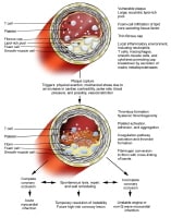 Pathogenesis of acute coronary syndromes. NextBackground
Chest pain is a nonspecific symptom that can have cardiac or noncardiac causes (see DDx). Unstable angina belongs to the spectrum of clinical presentations referred to collectively as acute coronary syndromes (ACSs), which range from ST-segment elevation myocardial infarction (STEMI) to non-STEMI (NSTEMI). Unstable angina is considered to be an ACS in which there is no detectable release of the enzymes and biomarkers of myocardial necrosis. The term angina is typically reserved for pain syndromes arising from presumed myocardial ischemia.
The traditional term unstable angina was meant to signify the intermediate state between myocardial infarction (MI) and the more chronic state of stable angina. The old term preinfarction angina conveys the clinical intent of intervening to attenuate the risk of MI or death. Patients with this condition have also been categorized by presentation, diagnostic test results, or course over time; these categories include new-onset angina, accelerating angina, rest angina, early postinfarct angina, and early postrevascularization angina.
Although the etiology and definition of unstable angina can be broad, interplay between disrupted atherosclerotic plaque and overlaid thrombi is present in many cases of unstable angina, with consequent hemodynamic deficit or microembolization. Thus, the condition is distinct from stable angina, in which the typical underlying cause is a fixed coronary stenosis with compromised blood flow and slow, progressive plaque growth that allows potential development of collateral vessels.
Other causes of angina, such as hypertrophic obstructive cardiomyopathy (HOCM) or microvascular disease (syndrome X), cause ischemia by means of different mechanisms and are considered separate entities.
PreviousNextPathophysiologyFactors involved in the pathophysiology of unstable angina include the following:
Supply-demand mismatchPlaque disruption or ruptureThrombosisVasoconstrictionCyclical flowSupply-demand mismatchThe myocardial ischemia of unstable angina, like all tissue ischemia, results from excessive demand or inadequate supply of oxygen, glucose, and free fatty acids.
Increased myocardial oxygen demand may be caused by the following:
FeverTachyarrhythmias (eg, atrial fibrillation or flutter)Malignant hypertensionThyrotoxicosisPheochromocytomaCocaine useAmphetamine useAortic stenosisSupravalvular aortic stenosisObstructive cardiomyopathyAortovenous shuntsHigh-output statesCongestive heart failure (CHF)Decreased oxygen supply may be caused by the following:
AnemiaHypoxemiaPolycythemiaHypotensionThe above causes must be investigated because a number of them are reversible. For example, anemia from chronic gastrointestinal (GI) bleeding is not uncommon in elderly patients. This can coexist with coronary artery disease (CAD). However, patients may not benefit from or may be harmed by treatments such as anticoagulants and antiplatelet drugs. Avoidance or treatment of the underlying condition is paramount.
Excess demand from increased myocardial workload (the product of heart rate and systolic blood pressure) or wall stress is responsible for nearly all cases of stable angina and perhaps one third of all episodes of unstable angina.
Plaque disruptionAccumulation of lipid-laden macrophages and smooth muscle cells, so-called foam cells, occurs within atherosclerotic plaques. The oxidized low-density lipoprotein cholesterol (LDL-C) in foam cells is cytotoxic, procoagulant, and chemotactic. As the atherosclerotic plaque grows, production of macrophage proteases and neutrophil elastases within the plaque can cause thinning of the fibromuscular cap that covers the lipid core.
Increasing plaque instability, coupled with blood-flow shear and circumferential wall stress, leads to plaque fissuring or rupture (see the image below), especially at the junction of the cap and the vessel wall. (See Vulnerable Plaque Pathology.)
Pathogenesis of acute coronary syndromes. The degree and consequences of plaque disruption cover a wide spectrum. Minor fissuring is typically nonocclusive and hence clinically silent, and repeat occult episodes of plaque ulceration and healing with a gradual growth of plaque volume have been histologically documented. Moderate-to-large plaque disruptions commonly result in unstable angina or acute infarction.
As many as 50% of MIs are due to lesions that are angiographically considered functionally insignificant.[2] Angiographically mild lesions can still be dangerous because they have an unstable thin-cap fibroatheroma (TCFA). This means that focal treatments such as percutaneous coronary intervention (PCI) are incomplete and that medical therapy to protect the entire vascular tree is complementary and crucial, particularly in patients with a history of ACS.
Vasoconstriction and thrombosisMost patients with ACS have recurrent transient reduction in coronary blood supply because of vasoconstriction and thrombus formation at the site of atherosclerotic plaque rupture. These events occur as consequences of episodic platelet aggregation and complex interactions among the vascular wall, leukocytes, platelets, and atherogenic lipoproteins.
Exposure of subendothelial components provokes platelet adhesion and activation. Platelets then aggregate in response to exposed vessel wall collagen or local aggregates (eg, thromboxane and adenosine diphosphate). Platelets also release substances that promote vasoconstriction and production of thrombin. In a reciprocating fashion, thrombin is a potent agonist for further platelet activation, and it stabilizes thrombi by converting fibrinogen to fibrin.
ACS may involve a clot in flux (ie, forming and enlarging, chipping off and embolizing). Over time, this dynamic clot formation or lysis, in conjunction with coronary vasoreactivity and resistance in the microvascular bed, causes intermittent and alternating (or cyclical) occlusion and flow.
The nonocclusive thrombus of unstable angina can become transiently or persistently occlusive. Depending on the duration of the occlusion, the presence of collateral vessels, and the area of myocardium perfused, recurrent unstable angina, non-Q-wave MI (NQMI), or Q-wave MI can result.
[#IntroductionEpidemiology] Genetics
Although the etiology of cardiovascular disease is strongly linked to modifiable environmental factors, it is known that genetics also play a significant part in the development of CAD and unstable angina. Much of the literature regarding the genetics of cardiovascular disease concerns MI and the development of CAD; however, there is a growing body of literature concerning unstable angina itself.
A number of genetic contributions are known to play a part in unstable angina. Genome-wide association studies (GWAS) have found linkage with unstable angina at chromosome 2q36-q37.3, chromosome 3q26-q27, and chromosome 20q11-13.[3] A polymorphism in glycoprotein Ia was associated with an increased time before platelet aggregation occurs in heterozygotes for the polymorphism in a Chinese population[4] ; it was postulated that the difference in platelet aggregation affected the pathogenesis of unstable angina.
Polymorphisms in several matrix metalloproteinase (MMP) genes have also been described. A guanine insertion in MMP1 is associated with smaller and more stable plaques, whereas the presence of more than 22 “CA†microsatellite repeats in MMP9 is associated with a worse prognosis for unstable angina.[5, 6]
Polymorphisms of interleukin (IL)-1 receptor antagonist (IL-1Ra) are suspected of having a role in the development of unstable angina. Studies conducted to date suggest that persons with allele-2 of IL-2Ra have increased inflammation, as measured by C-reactive protein (CRP) levels. There was an increased frequency of younger presentation in one study,[7] but a clear association between this polymorphism and an increased risk for unstable angina has not been found.
Apolipoprotein E (ApoE) polymorphisms also may play a pathogenetic role. In a study assessing the relation of ApoE4 to serum IL-10 levels, IL-10 levels were found to be lower in patients with at least 1 copy of ApoE4.[8] Higher IL-10 levels are believed to be cardioprotective, further suggesting that ApoE4 is associated with increased risk for unstable angina.[8] Ultimately, the genetics of unstable angina appear to be most closely linked with markers of inflammation and mediated by their effects on the risk of plaque rupture.[9]
PreviousNextEpidemiologyIn the United States, the incidence of unstable angina is increasing, and each year, nearly 1 million hospitalized patients have a primary diagnosis of unstable angina. A similar number of unstable angina episodes likely occur outside the hospital and either go unrecognized or are managed in the outpatient setting. However, even with heightened public awareness, improved survival after MI, and an aging population, the incidence of unstable angina should continue to rise despite primary and secondary prevention measures.
United States demographicsReasonably representative statistical estimates for unstable angina can be obtained from 2 registries, the GUARANTEE (Global Unstable Angina Registry and Treatment Evaluation) registry[10] and the CRUSADE (Can Rapid risk stratification of Unstable angina patients Suppress ADverse outcomes with Early implementation) registry of the American College of Cardiology (ACC) and the American Heart Association (AHA).[11, 12] (See Table 1 below.)
Table 1. Patient Characteristics, GUARANTEE Versus CRUSADE Trials (Open Table in a new window)
Characteristics GUARANTEE, 1995-96[10] CRUSADE, 2001-06[11] Mean age (y)6269Patients >65 y (%)44–Female (%)3940Hypertension (%)6073Diabetes mellitus (%)2633Current smoker (%)25–Hypercholesterolemia (%)4350Previous stroke (%)9–Previous MI (%)3630Previous angina (%)66–CHF (%)1418Previous coronary intervention (%)2321Previous coronary bypass surgery (%)2519CHF = congestive heart failure; CRUSADE = Can Rapid risk stratification of Unstable angina patients Suppress ADverse outcomes with Early implementation of the American College of Cardiology/American Heart Association guidelines; GUARANTEE = Global Unstable Angina Registry and Treatment Evaluation; MI = myocardial infarction.GUARANTEE involved 3000 consecutive hospital admissions for unstable angina in 35 US hospitals in 6 geographic regions (Northeast, Mideast, Midwest, Southeast, Southwest, Northwest) from September 1995 to August 1996.
CRUSADE registered more than 180,000 US patients with NSTEMI from 2001 to 2006, targeting high-risk patients with unstable angina or NSTEMI according to the following inclusion criteria, either separately or in combination[11, 12] :
Chest pain or anginal equivalent at rest, more than 10 minutes in durationIschemic electrocardiographic (ECG) changes (ST-segment depression >0.5 mm, transient ST-segment elevation 0.5-1.0 mm lasting for Elevated markers of myocardial necrosis (CK-MB or troponin I or T exceeding the upper limit of normal for the local laboratory assay used at each institution) International demographicsThe best international demographic data available are from the OASIS-2 (Organization to Assess Strategies for Ischemic Syndromes) registry (see Table 2 below).[13]
Table 2. Demographic Characteristics of Patients in International OASIS-2 Registry (Open Table in a new window)
Characteristics[13] Australia Brazil Canada Hungary Poland United States GeneralNumber of patients1899147816269311135918Mean age (y)656266656366Women (%)374237454037ClinicalNQMI presentation (%)7714221716Abnormal ECG (%)749182959787Select treatmentsBeta blocker (%)675373675957Calcium blocker (%)595153524359Invasive procedures (index hospitalization)Cardiac catheterization (%)24694320758PCI (%)7191650.424CABG (%)4201070.417CABG = coronary artery bypass grafting; ECG = electrocardiographic; NQMI = non-Q wave myocardial infarction; OASIS = Organization to Assess Strategies for Ischemic Syndromes; PCI = percutaneous coronary intervention.Because unstable angina is intimately linked to the incidence of coronary events, an approximation of international trends might be found in the MONICA (Monitoring Trends and Determinants in Cardiovascular Diseases) registry sponsored by the World Health Organization (WHO).[14] This large project monitored more than 7 million people aged 35-64 years from 30 populations in 21 countries from the mid-1980s.
In the study, the highest average rates of heart disease were found in Glasgow and Belfast, United Kingdom; North Karelia and Kuopio, Finland; Newcastle, Australia; and Warsaw, Poland.[14] The lowest average MI rates, and presumably the lowest average unstable angina rates, were observed in Beijing, China; Toulouse, France; Catalonia, Spain; Vaud-Fribourg, Switzerland; and Brianza, Italy.
The GRACE (Global Registry of Acute Coronary Events) registry (http://www.outcomes-umassmed.org/grace/) is prospectively tracking contemporary ACS treatment and outcome across 30 countries and has accumulated more than 100,000 patients.[15]
Age-related demographicsThe mean age of presentation with unstable angina is 62 years (range, 23-100 years). To put this in perspective, the mean age is 60 years for patients in clinical trials for MI, about 67 years for carotid artery stenosis, and 63 years for congestive heart failure. On average, women with unstable angina are 5 years older than men on presentation, with approximately half of women older than 65 years, as opposed to only about one third of men. Black individuals tend to present at a slightly younger age than people of other races do.
Sex-related demographicsWomen with unstable angina are older and have a higher prevalence of hypertension, diabetes mellitus, CHF, and family history of CAD than men. Men tend to have a higher previous incidence of MI and revascularization, a higher proportion of positive cardiac enzymes on admission, and higher rates of catheterization and revascularization. However, outcome is related more to the severity of the illness than to sex.
Race-related demographicsDisparities in outcome and risk-factor prevalence among different ethnic groups have been widely reported. For instance, as a group, black persons exhibit a higher prevalence of atherosclerotic risk factors (eg, hypertension, diabetes mellitus, and smoking), greater left ventricular mass, and decreased peripheral vasodilatory response. Relative to white persons, MI more frequently results in death in black individuals at young ages.
Fewer myocardial events but more cerebral complications have also been observed in black patients with unstable angina in randomized clinical trials of heparin versus hirudin (the Global Utilization of Streptokinase and TPA [tissue plasminogen activator] for Occluded coronary arteries II [GUSTO II] trial) or eptifibatide versus placebo (the Platelet glycoprotein IIb/IIIa in Unstable angina: Receptor Suppression Using Integrilin Therapy [PURSUIT] trial), possibly because of enhanced fibrinolytic activity and a higher prevalence of hypertension.
Racial differences also exist with regard to the delivery and response to medical care. White individuals have a higher rate of catheterization, angioplasty, and bypass surgery than individuals from other racial groups do.
Studies have shown equivalent short-term (30-day) mortality figures from unstable angina (including NQMI) for black individuals, but over the long term, persistent worse outcomes have been demonstrated.
PreviousNextPrognosisThe risk of MI, complications, and death in unstable angina varies because of the broad clinical spectrum that is covered by the term unstable angina. The aggressiveness of the therapeutic approach should be commensurate with the individualized estimated risk.
Patients who present with new ST-segment deviation (≥1 mm) have a 1-year death or MI rate of 11%, compared with a rate of only 6.8% in patients with isolated T-wave inversion.[16]
The current standard for cross-comparing studies is the 30-day event rate. The aggregate data for the more than 40,000 patients with ACSs (excluding STEMI), as derived from studies using contemporary treatments (albeit in varying degrees), indicate improving outcomes (see Table 3 below). The 30-day MI and death rates are currently around 8.5% and 3.5%, respectively, despite increased disease complexity and an aging cohort.
Table 3. Thirty-Day Clinical Outcome in Patients With Acute Coronary Syndromes in Clinical Trials (Open Table in a new window)
Study Year Number of Patients Death (%) Myocardial Infarction (%) Major Bleed (%) TIMI-319941,4732.59.00.3GUSTO-IIb19978,0113.86.01.0ESSENCE19983,1713.34.51.1PARAGON-A19982,2823.210.34.0PRISM19983,2323.04.20.4PRISM-PLUS19981,9154.48.11.1PURSUIT199810,9483.612.92.1TIMI-11B19993,9103.96.01.3PARAGON-B20005,2253.19.31.1Pooled40,1673.58.51.5ESSENCE = Efficacy and Safety of Subcutaneous Enoxaparin in Non–Q-wave Coronary Events; GUSTO-IIb = Global Utilization of Streptokinase and TPA (tissue plasminogen activator) for Occluded Coronary Arteries; PARAGON-A = Platelet IIb/IIIa Antagonism (lamifiban) for the Reduction of Acute Coronary Syndrome Events in a Global Organization Network; PARAGON-B = Platelet IIb/IIIa Antagonism (lamifiban) for the Reduction of Acute Coronary Syndrome Events in a Global Organization Network; PRISM = Platelet Receptor Inhibition in Ischemic Syndrome Management; PRISM-PLUS = Platelet Receptor Inhibition in Ischemic Syndrome Management in Patients Limited by Unstable Angina Signs and Symptoms; PURSUIT = Platelet Glycoprotein IIb/IIIa in Unstable Angina: Receptor Suppression Using Integrilin Therapy; TIMI-11B = Thrombolysis in Myocardial Infarction Clinical Trial 11B; TIMI-3 = Thrombolysis in Myocardial Infarction Clinical Trial 3.The RESCATE (Recursos Empleados en el Sindrome Coronario Agudo y Tiempos de Espera) investigators from Spain reported a 1.8% death rate and a 5.1% MI rate at 28 days (consecutive series, 1992-1994; early revascularization rate, ~6%) in 791 patients with unstable angina.[17] Compared with the rates in the North American studies listed earlier (see Epidemiology), these seem lower, probably because of the healthier case-mix; this illustrates the difficulties of direct outcome comparisons between institutions, countries, and trials.
A contemporary large clinical trial with centrally adjudicated outcomes showed that an ACS portends more adverse events in the year to come.[18] The following are the 12-month event rates for the ACS patients (final diagnosis of unstable angina, 16.6%; NSTEMI, 42.9%; STEMI, 37.5%), whose median age was 62 years, 25% of whom were diabetic, and fewer than 1% of whom were classified as above Killip class 2[18] :
Death from vascular causes - 4.3%Death from nonvascular causes - 0.6%MI - 5.9%Stroke - 1.2%These findings present an opportunity for secondary prevention of such adverse events.
Prognostic indicatorsOf note, studies have shown that the following are significant prognosticators for poor outcome in patients with unstable angina:
Ongoing CHFPresence or history of poor left ventricular ejection fraction (LVEF)Hemodynamic instabilityRecurrent angina despite intensive anti-ischemic therapyNew or worsening mitral regurgitationSustained ventricular tachycardiaAlthough these factors were not evaluated in the Thrombolysis in Myocardial Infarction (TIMI) Risk Score model (see Physical Examination), they should be taken into consideration when the level of care is decided.
Other predictors of worse long-term outcome in unstable angina include underlying left ventricular systolic dysfunction and more widespread extent of CAD.
The level of troponin positivity correlates with intermediate-term death in a dose-dependent fashion (range, 1.0-7.5% at 6 weeks) independent of age, creatine kinase MB isoenzyme (CK-MB) levels, and ST-segment deviation.
PreviousNextPatient EducationBefore hospital discharge, patients with unstable angina and their family members should be educated about the manifestations of MI and the actions that must to be taken in that eventuality. They should also receive training in cardiopulmonary resuscitation (CPR).
For patient information, see Cholesterol Center and Heart Health Center, as well as Cholesterol FAQs, Heart Disease FAQs, Angina, Chest Pain, High Cholesterol, Understanding Your Cholesterol level, Lifestyle CholesterolManagement, Understanding Cholesterol-Lowering Medications, and Heart Attack.
PreviousProceed to Clinical Presentation  Contributor Information and DisclosuresAuthorWalter Tan, MD, MS Director, Structural Heart Program, WakeMed Health and Hospitals; Specialist in Cardiovascular/Advanced Devices, Wake Specialty Physicians
Walter Tan, MD, MS is a member of the following medical societies: American Association for the Advancement of Science, American College of Cardiology, American Heart Association, American Stroke Association, National Stroke Association, Society for Vascular Medicine and Biology, and Society of Interventional Radiology
Disclosure: Nothing to disclose.
David J Moliterno, MD Professor of Medicine, Jefferson Morris Gill Professor of Cardiology, Chief, Division of Cardiovascular Medicine, University of Kentucky; Vice Chairman of Internal Medicine, Chandler Medical Center; Medical Director, Gill Heart Institute
David J Moliterno, MD is a member of the following medical societies: American College of Cardiology, American College of Physicians, American Heart Association, American Medical Association, Association of Professors of Cardiology, and European Society of Cardiology
Disclosure: Nothing to disclose.
Eric H Yang, MD Associate Professor of Medicine, Director of Cardiac Catherization Laboratory and Interventional Cardiology, Mayo Clinic Arizona
Eric H Yang, MD is a member of the following medical societies: Alpha Omega Alpha
Disclosure: Nothing to disclose.
Steven James Filby, MD Fellow in Interventional Cardiology, The Cleveland Clinic Foundation
Disclosure: Nothing to disclose.
Justin D Pearlman, MD, ME, PhD, FACC, MA Chief, Division of Cardiology, Director of Cardiology Consultative Service, Director of Cardiology Clinic Service, Director of Cardiology Non-Invasive Laboratory, Director of Cardiology Quality Program KMC, Dartmouth-Hitchcock Medical Center, Dartmouth Medical School
Justin D Pearlman, MD, ME, PhD, FACC, MA is a member of the following medical societies: American College of Cardiology, American College of Physicians, American Federation for Medical Research, International Society for Magnetic Resonance in Medicine, and Radiological Society of North America
Disclosure: Nothing to disclose.
Francisco Talavera, PharmD, PhD Adjunct Assistant Professor, University of Nebraska Medical Center College of Pharmacy; Editor-in-Chief, Medscape Drug Reference
Disclosure: Medscape Salary Employment
ReferenceseMedicineHealth. February 7, 2012. What are the symptoms of heart attack and unstable angina?. Available at http://www.emedicinehealth.com/heart_attack_and_unstable_angina-health/page5_em.htm#Symptoms.. Accessed May 9, 2013.
Stone GW, Maehara A, Lansky AJ, de Bruyne B, Cristea E, Mintz GS, et al. A prospective natural-history study of coronary atherosclerosis. N Engl J Med. Jan 20 2011;364(3):226-35. [Medline].
Harrap SB, Zammit KS, Wong ZY, Williams FM, Bahlo M, Tonkin AM, et al. Genome-wide linkage analysis of the acute coronary syndrome suggests a locus on chromosome 2. Arterioscler Thromb Vasc Biol. May 1 2002;22(5):874-8. [Medline].
Zhao YH, Xu Y, Gu YY, Li Y, Zhang JY, Su X. Functional effect of platelet membrane glycoprotein ia gene polymorphism in the pathogenesis of unstable angina pectoris. J Int Med Res. 2011;39(2):541-8. [Medline].
Fiotti N, Moretti ME, Bussani R, Altamura N, Zamolo F, Gerloni R, et al. Features of vulnerable plaques and clinical outcome of UA/NSTEMI: Relationship with matrix metalloproteinase functional polymorphisms. Atherosclerosis. Mar 2011;215(1):153-9. [Medline].
White AJ, Duffy SJ, Walton AS, Ng JF, Rice GE, Mukherjee S, et al. Matrix metalloproteinase-3 and coronary remodelling: implications for unstable coronary disease. Cardiovasc Res. Sep 1 2007;75(4):813-20. [Medline].
Manzoli A, Andreotti F, Varlotta C, Mollichelli N, Verde M, van de Greef W, et al. Allelic polymorphism of the interleukin-1 receptor antagonist gene in patients with acute or stable presentation of ischemic heart disease. Cardiologia. Sep 1999;44(9):825-30. [Medline].
Tziakas DN, Chalikias GK, Antonoglou CO, Veletza S, Tentes IK, Kortsaris AX, et al. Apolipoprotein E genotype and circulating interleukin-10 levels in patients with stable and unstable coronary artery disease. J Am Coll Cardiol. Dec 19 2006;48(12):2471-81. [Medline].
Willerson JT. Systemic and local inflammation in patients with unstable atherosclerotic plaques. Prog Cardiovasc Dis. May-Jun 2002;44(6):469-78. [Medline].
Scirica BM, Moliterno DJ, Every NR, Anderson HV, Aguirre FV, Granger CB, et al. Differences between men and women in the management of unstable angina pectoris (The GUARANTEE Registry). The GUARANTEE Investigators. Am J Cardiol. Nov 15 1999;84(10):1145-50. [Medline].
Skolnick AH, Alexander KP, Chen AY, Roe MT, Pollack CV Jr, Ohman EM, et al. Characteristics, management, and outcomes of 5,557 patients age > or =90 years with acute coronary syndromes: results from the CRUSADE Initiative. J Am Coll Cardiol. May 1 2007;49(17):1790-7. [Medline].
Hoekstra JW, Pollack CV Jr, Roe MT, Peterson ED, Brindis R, Harrington RA, et al. Improving the care of patients with non-ST-elevation acute coronary syndromes in the emergency department: the CRUSADE initiative. Acad Emerg Med. Nov 2002;9(11):1146-55. [Medline].
Effects of recombinant hirudin (lepirudin) compared with heparin on death, myocardial infarction, refractory angina, and revascularisation procedures in patients with acute myocardial ischaemia without ST elevation: a randomised trial. Organisation to Assess Strategies for Ischemic Syndromes (OASIS-2) Investigators. Lancet. Feb 6 1999;353(9151):429-38. [Medline].
Luepker RV. WHO MONICA project: what have we learned and where to go from here?. Public Health Rev. 2012. Accessed: May 8, 2013;33(2):373-96. [Medline].
GRACE, Global Registry of Acute Coronary Events. Available at http://www.outcomes-umassmed.org/grace/. Accessed September 16, 2010.
Cannon CP, McCabe CH, Stone PH, Rogers WJ, Schactman M, Thompson BW, et al. The electrocardiogram predicts one-year outcome of patients with unstable angina and non-Q wave myocardial infarction: results of the TIMI III Registry ECG Ancillary Study. Thrombolysis in Myocardial Ischemia. J Am Coll Cardiol. Jul 1997;30(1):133-40. [Medline].
Lupón J, Valle V, Marrugat J, Elosua R, Serés L, Pavesi M, et al. Six-month outcome in unstable angina patients without previous myocardial infarction according to the use of tertiary cardiologic resources. RESCATE Investigators. Recursos Empleados en el SÃndrome Coronario Agudo y Tiempos de Espera. J Am Coll Cardiol. Dec 1999;34(7):1947-53. [Medline].
Wallentin L, Becker RC, Budaj A, Cannon CP, Emanuelsson H, Held C, et al. Ticagrelor versus clopidogrel in patients with acute coronary syndromes. N Engl J Med. Sep 10 2009;361(11):1045-57. [Medline].
Karcz A, Holbrook J, Burke MC, Doyle MJ, Erdos MS, Friedman M, et al. Massachusetts emergency medicine closed malpractice claims: 1988-1990. Ann Emerg Med. Mar 1993;22(3):553-9. [Medline].
Meune C, Balmelli C, Twerenbold R, Reichlin T, Reiter M, Haaf P, et al. Patients with acute coronary syndrome and normal high-sensitivity troponin. Am J Med. Dec 2011;124(12):1151-7. [Medline].
Misra D, Leibowitz K, Gowda RM, Shapiro M, Khan IA. Role of N-acetylcysteine in prevention of contrast-induced nephropathy after cardiovascular procedures: a meta-analysis. Clin Cardiol. Nov 2004;27(11):607-10. [Medline].
Than M, Cullen L, Reid CM, Lim SH, Aldous S, Ardagh MW, et al. A 2-h diagnostic protocol to assess patients with chest pain symptoms in the Asia-Pacific region (ASPECT): a prospective observational validation study. Lancet. Mar 26 2011;377(9771):1077-84. [Medline].
Thygesen K, Alpert JS, Jaffe AS, Simoons ML, Chaitman BR, White HD. Third universal definition of myocardial infarction. Eur Heart J. Oct 2012;33(20):2551-67. [Medline]. [Full Text].
Januzzi JL, Cannon CP, DiBattiste PM, Murphy S, Weintraub W, Braunwald E. Effects of renal insufficiency on early invasive management in patients with acute coronary syndromes (The TACTICS-TIMI 18 Trial). Am J Cardiol. Dec 1 2002;90(11):1246-9. [Medline].
Acute coronary syndrome and myocardial infarction. In: EBM Guidelines. Evidence-Based Medicine [internet]. Helsinki, Finland: Wiley Interscience. John Wiley & Sons; 2011:[Full Text].
de Zwaan C, Bar FW, Wellens HJ. Characteristic electrocardiographic pattern indicating a critical stenosis high in left anterior descending coronary artery in patients admitted because of impending myocardial infarction. Am Heart J. Apr 1982;;103(4 Pt 2):730-6. [Medline].
Nisbet BC, Zlupko G. Repeat Wellens' syndrome: case report of critical proximal left anterior descending artery restenosis. J Emerg Med. Sep 2010;39(3):305-8. [Medline].
Kwong RY, Chan AK, Brown KA, et al. Impact of unrecognized myocardial scar detected by cardiac magnetic resonance imaging on event-free survival in patients presenting with signs or symptoms of coronary artery disease. Circulation. Jun 13 2006;113(23):2733-43. [Medline].
Kwong RY, Sattar H, Wu H, et al. Incidence and prognostic implication of unrecognized myocardial scar characterized by cardiac magnetic resonance in diabetic patients without clinical evidence of myocardial infarction. Circulation. Sep 2 2008;118(10):1011-20. [Medline]. [Full Text].
Stratmann HG, Younis LT, Wittry MD, Amato M, Miller DD. Exercise technetium-99m myocardial tomography for the risk stratification of men with medically treated unstable angina pectoris. Am J Cardiol. Aug 1 1995;76(4):236-40. [Medline].
Udelson JE, Spiegler EJ. Emergency department perfusion imaging for suspected coronary artery disease: the ERASE Chest Pain Trial. Md Med. Spring 2001;Suppl:90-4. [Medline].
[Guideline] Wright RS, Anderson JL, Adams CD, Bridges CR, Casey DE Jr, Ettinger SM, et al. 2011 ACCF/AHA Focused Update of the Guidelines for the Management of Patients With Unstable Angina/Non-ST-Elevation Myocardial Infarction (Updating the 2007 Guideline): A Report of the American College of Cardiology Foundation/American Heart Association Task Force on Practice Guidelines. Circulation. Mar 28 2011;[Medline].
Yusuf S, Zhao F, Mehta SR, Chrolavicius S, Tognoni G, Fox KK. Effects of clopidogrel in addition to aspirin in patients with acute coronary syndromes without ST-segment elevation. N Engl J Med. Aug 16 2001;345(7):494-502. [Medline].
Peters RJ, Mehta SR, Fox KA, Zhao F, Lewis BS, Kopecky SL, et al. Effects of aspirin dose when used alone or in combination with clopidogrel in patients with acute coronary syndromes: observations from the Clopidogrel in Unstable angina to prevent Recurrent Events (CURE) study. Circulation. Oct 7 2003;108(14):1682-7. [Medline].
Steinhubl SR, Berger PB, Mann JT 3rd, Fry ET, DeLago A, Wilmer C, et al. Early and sustained dual oral antiplatelet therapy following percutaneous coronary intervention: a randomized controlled trial. JAMA. Nov 20 2002;288(19):2411-20. [Medline].
Mega JL, Close SL, Wiviott SD, Shen L, Hockett RD, Brandt JT, et al. Cytochrome p-450 polymorphisms and response to clopidogrel. N Engl J Med. Jan 22 2009;360(4):354-62. [Medline].
Paré G, Mehta SR, Yusuf S, Anand SS, Connolly SJ, Hirsh J, et al. Effects of CYP2C19 genotype on outcomes of clopidogrel treatment. N Engl J Med. Oct 28 2010;363(18):1704-14. [Medline].
Park KW, Kim HS. Options to overcome clopidogrel response variability. Circ J. 2012;76(2):287-92. [Medline].
O'Connor FF, Shields DC, Fitzgerald A, Cannon CP, Braunwald E, Fitzgerald DJ. Genetic variation in glycoprotein IIb/IIIa (GPIIb/IIIa) as a determinant of the responses to an oral GPIIb/IIIa antagonist in patients with unstable coronary syndromes. Blood. Dec 1 2001;98(12):3256-60. [Medline].
National Clinical Guideline Centre for Acute and Chronic Conditions. Unstable angina and NSTEMI: the early management of unstable angina and non-ST-segment-elevation myocardial infarction. London, UK: National Institute for Health and Clinical Excellence; 2010:[Full Text].
Jneid H, Anderson JL, Wright RS, Adams CD, Bridges CR, Casey DE Jr, et al. 2012 ACCF/AHA focused update of the guideline for the management of patients with unstable angina/non-ST-elevation myocardial infarction (updating the 2007 guideline and replacing the 2011 focused update): a report of the American College of Cardiology Foundation/American Heart Association Task Force on Practice Guidelines. J Am Coll Cardiol. Aug 14 2012;60(7):645-81. [Medline].
Simoons ML. Effect of glycoprotein IIb/IIIa receptor blocker abciximab on outcome in patients with acute coronary syndromes without early coronary revascularisation: the GUSTO IV-ACS randomised trial. Lancet. Jun 16 2001;357(9272):1915-24. [Medline].
Ibbotson T, McGavin JK, Goa KL. Abciximab: an updated review of its therapeutic use in patients with ischaemic heart disease undergoing percutaneous coronary revascularisation. Drugs. 2003;63(11):1121-63. [Medline].
Roffi M, Chew DP, Mukherjee D, Bhatt DL, White JA, Heeschen C, et al. Platelet glycoprotein IIb/IIIa inhibitors reduce mortality in diabetic patients with non-ST-segment-elevation acute coronary syndromes. Circulation. Dec 4 2001;104(23):2767-71. [Medline].
Cohen M, Demers C, Gurfinkel EP, Turpie AG, Fromell GJ, Goodman S, et al. Low-molecular-weight heparins in non-ST-segment elevation ischemia: the ESSENCE trial. Efficacy and Safety of Subcutaneous Enoxaparin versus intravenous unfractionated heparin, in non-Q-wave Coronary Events. Am J Cardiol. Sep 10 1998;82(5B):19L-24L. [Medline].
Ferguson JJ, Califf RM, Antman EM, Cohen M, Grines CL, Goodman S, et al. Enoxaparin vs unfractionated heparin in high-risk patients with non-ST-segment elevation acute coronary syndromes managed with an intended early invasive strategy: primary results of the SYNERGY randomized trial. JAMA. Jul 7 2004;292(1):45-54. [Medline].
Mehta SR, Granger CB, Eikelboom JW, Bassand JP, Wallentin L, Faxon DP, et al. Efficacy and safety of fondaparinux versus enoxaparin in patients with acute coronary syndromes undergoing percutaneous coronary intervention: results from the OASIS-5 trial. J Am Coll Cardiol. Oct 30 2007;50(18):1742-51. [Medline].
Théroux P, Waters D, Lam J, Juneau M, McCans J. Reactivation of unstable angina after the discontinuation of heparin. N Engl J Med. Jul 16 1992;327(3):141-5. [Medline].
Direct thrombin inhibitors in acute coronary syndromes: principal results of a meta-analysis based on individual patients' data. Lancet. Jan 26 2002;359(9303):294-302. [Medline].
Metz BK, White HD, Granger CB, Simes RJ, Armstrong PW, Hirsh J, et al. Randomized comparison of direct thrombin inhibition versus heparin in conjunction with fibrinolytic therapy for acute myocardial infarction: results from the GUSTO-IIb Trial. Global Use of Strategies to Open Occluded Coronary Arteries in Acute Coronary Syndromes (GUSTO-IIb) Investigators. J Am Coll Cardiol. Jun 1998;31(7):1493-8. [Medline].
Maroo A, Lincoff AM. Bivalirudin in PCI: an overview of the REPLACE-2 trial. Semin Thromb Hemost. Jun 2004;30(3):329-36. [Medline].
Stone GW, McLaurin BT, Cox DA, Bertrand ME, Lincoff AM, Moses JW, et al. Bivalirudin for patients with acute coronary syndromes. N Engl J Med. Nov 23 2006;355(21):2203-16. [Medline].
Schwartz GG, Olsson AG, Ezekowitz MD, Ganz P, Oliver MF, Waters D, et al. Effects of atorvastatin on early recurrent ischemic events in acute coronary syndromes: the MIRACL study: a randomized controlled trial. JAMA. Apr 4 2001;285(13):1711-8. [Medline].
Murphy SA, Cannon CP, Wiviott SD, McCabe CH, Braunwald E. Reduction in recurrent cardiovascular events with intensive lipid-lowering statin therapy compared with moderate lipid-lowering statin therapy after acute coronary syndromes from the PROVE IT-TIMI 22 (Pravastatin or Atorvastatin Evaluation and Infection Therapy-Thrombolysis In Myocardial Infarction 22) trial. J Am Coll Cardiol. Dec 15 2009;54(25):2358-62. [Medline].
US Food and Drug Administration. Safety: statin drugs - drug safety communication: class labeling change. Posted: February 28, 2012. Available at http://www.fda.gov/Safety/MedWatch/SafetyInformation/SafetyAlertsforHumanMedicalProducts/ucm293670.htm. Accessed June 5, 2013.
US Food and Drug Administration. Safety: Zocor (simvastatin): label change - new restrictions, contraindications, and dose limitations. Posted: June 8, 2011. Available at http://www.fda.gov/Safety/MedWatch/SafetyInformation/SafetyAlertsforHumanMedicalProducts/ucm258384.htm. Accessed June 5, 2013.
US Food and Drug Administration. Safety: Meridia (sibutramine): market withdrawal due to risk of serious cardiovascular events. Posted: October 8, 2010. Available at http://www.fda.gov/Safety/MedWatch/SafetyInformation/SafetyAlertsforHumanMedicalProducts/ucm228830.htm. Accessed June 5, 2013.
Anderson HV, Cannon CP, Stone PH, Williams DO, McCabe CH, Knatterud GL, et al. One-year results of the Thrombolysis in Myocardial Infarction (TIMI) IIIB clinical trial. A randomized comparison of tissue-type plasminogen activator versus placebo and early invasive versus early conservative strategies in unstable angina and non-Q wave myocardial infarction. J Am Coll Cardiol. Dec 1995;26(7):1643-50. [Medline].
Boden WE, O'Rourke RA, Crawford MH, Blaustein AS, Deedwania PC, Zoble RG. Outcomes in patients with acute non-Q-wave myocardial infarction randomly assigned to an invasive as compared with a conservative management strategy. Veterans Affairs Non-Q-Wave Infarction Strategies in Hospital (VANQWISH) Trial Investigators. N Engl J Med. Jun 18 1998;338(25):1785-92. [Medline].
Invasive compared with non-invasive treatment in unstable coronary-artery disease: FRISC II prospective randomised multicentre study. FRagmin and Fast Revascularisation during InStability in Coronary artery disease Investigators. Lancet. Aug 28 1999;354(9180):708-15. [Medline].
Fox KA, Poole-Wilson PA, Henderson RA, Clayton TC, Chamberlain DA, Shaw TR, et al. Interventional versus conservative treatment for patients with unstable angina or non-ST-elevation myocardial infarction: the British Heart Foundation RITA 3 randomised trial. Randomized Intervention Trial of unstable Angina. Lancet. Sep 7 2002;360(9335):743-51. [Medline].
Damman P, Hirsch A, Windhausen F, Tijssen JG, de Winter RJ. 5-year clinical outcomes in the ICTUS (Invasive versus Conservative Treatment in Unstable coronary Syndromes) trial a randomized comparison of an early invasive versus selective invasive management in patients with non-ST-segment elevation acute coronary syndrome. J Am Coll Cardiol. Mar 2 2010;55(9):858-64. [Medline].
Neumann FJ, Kastrati A, Pogatsa-Murray G, Mehilli J, Bollwein H, Bestehorn HP, et al. Evaluation of prolonged antithrombotic pretreatment ("cooling-off" strategy) before intervention in patients with unstable coronary syndromes: a randomized controlled trial. JAMA. Sep 24 2003;290(12):1593-9. [Medline].
[Guideline] Anderson JL, Adams CD, Antman EM, Bridges CR, Califf RM, Casey DE Jr, et al. ACC/AHA 2007 guidelines for the management of patients with unstable angina/non ST-elevation myocardial infarction: a report of the American College of Cardiology/American Heart Association Task Force on Practice Guidelines (Writing Committee to Revise the 2002 Guidelines for the Management of Patients With Unstable Angina/Non ST-Elevation Myocardial Infarction): developed in collaboration with the American College of Emergency Physicians, the Society for Cardiovascular Angiography and Interventions, and the Society of Thoracic Surgeons: endorsed by the American Association of Cardiovascular and Pulmonary Rehabilitation and the Society for Academic Emergency Medicine. Circulation. Aug 14 2007;116(7):e148-304. [Medline].
Wallentin L, Lagerqvist B, Husted S, Kontny F, Ståhle E, Swahn E. Outcome at 1 year after an invasive compared with a non-invasive strategy in unstable coronary-artery disease: the FRISC II invasive randomised trial. FRISC II Investigators. Fast Revascularisation during Instability in Coronary artery disease. Lancet. Jul 1 2000;356(9223):9-16. [Medline].
[Guideline] American Diabetes Association. Standards of medical care in diabetes--2010. Diabetes Care. Jan 2010;33 Suppl 1:S11-61. [Medline]. [Full Text].
A randomised, blinded, trial of clopidogrel versus aspirin in patients at risk of ischaemic events (CAPRIE). CAPRIE Steering Committee. Lancet. Nov 16 1996;348(9038):1329-39. [Medline].
Kastrati A, Neumann FJ, Schulz S, et al. Abciximab and heparin versus bivalirudin for non-ST-elevation myocardial infarction. N Engl J Med. Nov 24 2011;365(21):1980-9. [Medline].
Ong P, Athanasiadis A, Hill S, Vogelsberg H, Voehringer M, Sechtem U. Coronary artery spasm as a frequent cause of acute coronary syndrome: The CASPAR (Coronary Artery Spasm in Patients With Acute Coronary Syndrome) Study. J Am Coll Cardiol. Aug 12 2008;52(7):523-7. [Medline].
Â Pathogenesis of acute coronary syndromes. Thrombolysis in Myocardial Infarction (TIMI) Risk Score correlates with major adverse outcome and effect of therapy with low-molecular-weight heparin. ARD = absolute risk difference; ESSENCE = Efficacy and Safety of Subcutaneous Enoxaparin in Non–Q-wave Coronary Events; No. = number; NNT = number needed to treat. Algorithm for initial invasive strategy. ASA = acetylsalicylic acid (aspirin); GP IIb/IIIa= glycoprotein IIb/IIIa; IV = intravenous; LOE = level of evidence; UA/NSTEMI = unstable angina/non–ST-segment elevation myocardial infarction; UFH = unfractionated heparin. (Adapted from 2007 ACC/AHA UA/NSTEMI Guidelines.) Algorithm for initial conservative strategy. ASA = acetylsalicylic acid (aspirin); EF = ejection fraction; GP IIb/IIIa= glycoprotein IIb/IIIa; IV = intravenous; LOE = level of evidence; LVEF = left ventricular ejection fraction; UA/NSTEMI = unstable angina/non–ST-segment elevation myocardial infarction. (Adapted from 2007 ACC/AHA UA/NSTEMI Guidelines.) Rate and timing of revascularization for patients with unstable angina using invasive versus conservative approach (FRagmin during InStability in Coronary artery disease [FRISC] II). Time course of elevations of serum markers after acute myocardial infarction. CK = creatine kinase; CK-MB = creatine kinase MB fraction; LDH = lactate dehydrogenase. Table 1. Patient Characteristics, GUARANTEE Versus CRUSADE TrialsTable 2. Demographic Characteristics of Patients in International OASIS-2 RegistryTable 3. Thirty-Day Clinical Outcome in Patients With Acute Coronary Syndromes in Clinical TrialsTable 4. Braunwald Classification of Unstable AnginaTable 5. ACC/AHA Recommendations for Preferred Invasive StrategyTable 1. Patient Characteristics, GUARANTEE Versus CRUSADE TrialsCharacteristics GUARANTEE, 1995-96[10] CRUSADE, 2001-06[11] Mean age (y)6269Patients >65 y (%)44–Female (%)3940Hypertension (%)6073Diabetes mellitus (%)2633Current smoker (%)25–Hypercholesterolemia (%)4350Previous stroke (%)9–Previous MI (%)3630Previous angina (%)66–CHF (%)1418Previous coronary intervention (%)2321Previous coronary bypass surgery (%)2519CHF = congestive heart failure; CRUSADE = Can Rapid risk stratification of Unstable angina patients Suppress ADverse outcomes with Early implementation of the American College of Cardiology/American Heart Association guidelines; GUARANTEE = Global Unstable Angina Registry and Treatment Evaluation; MI = myocardial infarction. Table 2. Demographic Characteristics of Patients in International OASIS-2 RegistryCharacteristics[13] Australia Brazil Canada Hungary Poland United States GeneralNumber of patients1899147816269311135918Mean age (y)656266656366Women (%)374237454037ClinicalNQMI presentation (%)7714221716Abnormal ECG (%)749182959787Select treatmentsBeta blocker (%)675373675957Calcium blocker (%)595153524359Invasive procedures (index hospitalization)Cardiac catheterization (%)24694320758PCI (%)7191650.424CABG (%)4201070.417CABG = coronary artery bypass grafting; ECG = electrocardiographic; NQMI = non-Q wave myocardial infarction; OASIS = Organization to Assess Strategies for Ischemic Syndromes; PCI = percutaneous coronary intervention. Table 3. Thirty-Day Clinical Outcome in Patients With Acute Coronary Syndromes in Clinical TrialsStudy Year Number of Patients Death (%) Myocardial Infarction (%) Major Bleed (%) TIMI-319941,4732.59.00.3GUSTO-IIb19978,0113.86.01.0ESSENCE19983,1713.34.51.1PARAGON-A19982,2823.210.34.0PRISM19983,2323.04.20.4PRISM-PLUS19981,9154.48.11.1PURSUIT199810,9483.612.92.1TIMI-11B19993,9103.96.01.3PARAGON-B20005,2253.19.31.1Pooled40,1673.58.51.5ESSENCE = Efficacy and Safety of Subcutaneous Enoxaparin in Non–Q-wave Coronary Events; GUSTO-IIb = Global Utilization of Streptokinase and TPA (tissue plasminogen activator) for Occluded Coronary Arteries; PARAGON-A = Platelet IIb/IIIa Antagonism (lamifiban) for the Reduction of Acute Coronary Syndrome Events in a Global Organization Network; PARAGON-B = Platelet IIb/IIIa Antagonism (lamifiban) for the Reduction of Acute Coronary Syndrome Events in a Global Organization Network; PRISM = Platelet Receptor Inhibition in Ischemic Syndrome Management; PRISM-PLUS = Platelet Receptor Inhibition in Ischemic Syndrome Management in Patients Limited by Unstable Angina Signs and Symptoms; PURSUIT = Platelet Glycoprotein IIb/IIIa in Unstable Angina: Receptor Suppression Using Integrilin Therapy; TIMI-11B = Thrombolysis in Myocardial Infarction Clinical Trial 11B; TIMI-3 = Thrombolysis in Myocardial Infarction Clinical Trial 3. Table 4. Braunwald Classification of Unstable AnginaCharacteristic Class/Category Details SeverityISymptoms with exertionIISubacute symptoms at rest (2-30 days prior)IIIAcute symptoms at rest (within prior 48 hr)Clinical precipitating factorASecondaryBPrimaryCPostinfarctionTherapy during symptoms1No treatment2Usual angina therapy3Maximal therapyTable 5. ACC/AHA Recommendations for Preferred Invasive StrategyPreferred Strategy[64] Patient Characteristics InvasiveRecurrent angina/ischemia at rest or with low-level activities despite intensive medical therapyElevated cardiac biomarkers (TnT or TnI)New or presumably new ST-segment depressionSigns or symptoms of heart failure or new or worsening mitral regurgitationHigh-risk findings on noninvasive stress testingHigh-risk score (eg, TIMI, GRACE)Reduced LV systolic function (LVEF Hemodynamic instabilitySustained ventricular tachycardiaPCI within 6 monthsPrevious CABGConservativeLow-risk score (eg, TIMI, GRACE)Patient or physician preference in the absence of high-risk featuresACC/AHA = American College of Cardiology/American Heart Association; CABG = coronary artery bypass grafting; GRACE = Global Registry of Acute Coronary Events; LV = left ventricle; LVEF = left ventricular ejection fraction; PCI = percutaneous coronary intervention; TIMI = Thrombolysis in Myocardial Infarction Clinical Trial; TnI = troponin I; TnT = troponin T.  View Table List Read more about Unstable Angina on MedscapeRelated Reference Topics
Wellens Syndrome
Coronary Bare-Metal Stent
Percutaneous Coronary Intervention
Related News and Articles
2012 ACCF/AHA Focused Update of the Guideline for the Management of Patients With Unstable Angina/Non–ST-Elevation Myocardial Infarction (Updating the 2007 Guideline and Replacing the 2011 Focused Update)
What Do Angina Patients Understand of Options for Myocardial Revascularisation?
Changes of Serum Angiogenic Factors Concentrations in Patients With Diabetes and Unstable Angina Pectoris
Medscape Reference © 2011 WebMD, LLC, Unstable Angina


I would like to say that this blog really convinced me to do it! Thanks, very good post. Pancreatic cancer treatment
ReplyDelete