Superior vena cava syndrome (SVCS) is obstruction of blood flow through the superior vena cava (SVC). It is a medical emergency and most often manifests in patients with a malignant disease process within the thorax. A patient with superior vena cava syndrome (SVCS) requires immediate diagnostic evaluation and therapy. See the images below.
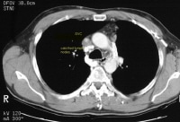 Superior vena cava syndrome (case 1). The patient was a 35-year-old man with a 3-year history of progressive upper-extremity and fascial swelling. The patient had undergone treatment for histoplasmosis in the past. CT scan shows a narrowed superior vena cava with adjacent calcified lymph nodes and posterior soft tissue thickening.
 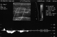 Superior vena cava syndrome (case 1, cont'd). Sonogram shows markedly damped venous waveform with complete loss of normal venous pulsatility and minimal respiratory variation.
 William Hunter first described the syndrome in 1757 in a patient with syphilitic aortic aneurysm.[1] In 1954, Schechter reviewed 274 well-documented cases of superior vena cava syndrome (SVCS) reported in the literature; 40% of them were due to syphilitic aneurysms or tuberculous mediastinitis.[2] In more recent times, these infections have gradually decreased as the primary cause of superior vena cava (SVC) obstruction. Lung cancer, particularly adenocarcinoma, is now the underlying process in approximately 70% of the patients with superior vena cava syndrome (SVCS).[3, 4, 5] However, up to 40% of the causes are due to nonmalignant causes.[6]
NextPathophysiologyThe superior vena cava (SVC) is the major drainage vessel for venous blood from the head, neck, upper extremities, and upper thorax. It is located in the middle mediastinum and is surrounded by relatively rigid structures such as the sternum, trachea, right bronchus, aorta, pulmonary artery, and the perihilar and paratracheal lymph nodes. It extends from the junction of the right and left innominate veins to the right atrium, a distance of 6-8 cm. It is a thin-walled, low-pressure, vascular structure. This wall is easily compressed as it traverses the right side of the mediastinum.[7]
Obstruction of the superior vena cava (SVC) may be caused by neoplastic invasion of the venous wall associated with intravascular thrombosis or, more simply, by extrinsic pressure of a tumor mass against the relatively fixed thin-walled superior vena cava (SVC). Postmortem examinations reveal that complete superior vena cava (SVC) obstruction is the result of intravascular thrombosis in combination with extrinsic pressure. Incomplete superior vena cava (SVC) obstruction is more often secondary to extrinsic compression without thrombosis. Other causes include compression by intravascular arterial devices. The incidence is on the rise in line with the increased usage of endovascular devices.[4]
An obstructed superior vena cava (SVC) initiates collateral venous return to the heart from the upper half of the body through 4 principal pathways. The first and most important pathway is the azygous venous system, which includes the azygous vein, the hemiazygous vein, and the connecting intercostal veins. The second pathway is the internal mammary venous system plus tributaries and secondary communications to the superior and inferior epigastric veins. The long thoracic venous system, with its connections to the femoral veins and vertebral veins, provides the third and fourth collateral routes, respectively.
Despite these collateral pathways, venous pressure is almost always elevated in the upper compartment if obstruction of the superior vena cava (SVC) is present. Venous pressure as high as 200-500 cm H2 O has been recorded in patients with severe superior vena cava syndrome (SVCS).
PreviousNextEpidemiologyFrequencyUnited StatesSuperior vena cava syndrome (SVCS) develops in 5-10% of patients with a right-sided malignant intrathoracic mass lesion. In 1969, Salsali and Cliffton observed superior vena cava syndrome (SVCS) in 4.2% of 4960 patients with lung cancer; 80% of the tumors inducing superior vena cava syndrome (SVCS) were of the right lung.[8] In 5 large series of small cell lung cancer, 9-19% of patients demonstrated superior vena cava syndrome (SVCS). In 1987, Armstrong and Perez found superior vena cava syndrome (SVCS) in 1.9% of 952 patients with lymphoma.[9]
Mortality/MorbiditySurvival in patients with superior vena cava syndrome (SVCS) depends mainly on the course of the underlying disease. No mortality, per se, results directly from mild venous congestion. In patients with benign superior vena cava syndrome (SVCS), life expectancy is unchanged.If superior vena cava syndrome (SVCS) is secondary to a malignant process, patient survival correlates with the histology of the tumor. Patients with signs and symptoms of laryngeal and cerebral edema have the most life-threatening manifestations of this syndrome and are in danger of sudden death. Clinical observations show that approximately 10% of patients with a bronchogenic carcinoma and 45% of patients with lymphoma treated with irradiation live at least 30 months. In contrast, patients with untreated malignant superior vena cava syndrome (SVCS) survive only approximately 30 days.[5] RaceThe frequency of superior vena cava syndrome (SVCS) in different races depends largely on the frequency of lung cancer and lymphomas in these populations.
SexMalignant causes of superior vena cava syndrome (SVCS) are most commonly observed in males because of the high incidence of lung cancer in this population. In contrast, no sex difference is observed in cases related to benign causes.AgeMalignant causes of superior vena cava syndrome (SVCS) are predominantly observed in individuals aged 40-60 years.Benign causes account for most of the cases diagnosed in individuals aged 30-40 years.Obstruction of the superior vena cava (SVC) in the pediatric age group is rare and has a different etiologic spectrum.PreviousProceed to Clinical Presentation , Superior Vena Cava Syndrome


0 comments:
Post a Comment