Infective endocarditis (IE) is defined as an infection of the endocardial surface of the heart, which may include one or more heart valves, the mural endocardium, or a septal defect. Its intracardiac effects include severe valvular insufficiency, which may lead to intractable congestive heart failure and myocardial abscesses. If left untreated, IE is generally fatal.
Essential update: Ceftriaxone an alternative treatment for infective endocarditisIn a recent study, the combination of ampicillin and ceftriaxone was shown to be as effective as the combination of ampicillin and gentamicin for treating Enterococcus faecalis infective endocarditis. In this observational study, mortality rates did not differ significantly between the 2 treatments either during antimicrobial therapy or at 3-month follow-up. There were also no significant differences in treatment failures resulting in a change of antimicrobials or in disease relapses.[1, 2]
According to study findings, the need to interrupt antibiotic treatment as a result of adverse events occurred significantly more often in patients treated with the ampicillin/gentamicin combination, primarily because of newly developed renal failure.[1, 2]
Signs and symptomsFever, possibly low-grade and intermittent, is present in 90% of patients with IE. Heart murmurs are heard in approximately 85% of patients.
One or more classic signs of IE are found in as many as 50% of patients. They include the following:
Petechiae: Common, but nonspecific, findingSubungual (splinter) hemorrhages: Dark-red, linear lesions in the nail bedsOsler nodes: Tender subcutaneous nodules usually found on the distal pads of the digitsJaneway lesions: Nontender maculae on the palms and solesRoth spots: Retinal hemorrhages with small, clear centers; rareSigns of neurologic disease, which occur in as many as 40% of patients, include the following[3] :
Embolic stroke with focal neurologic deficits: The most common neurologic signIntracerebral hemorrhageMultiple microabscessesOther signs of IE include the following:
SplenomegalyStiff neckDeliriumParalysis, hemiparesis, aphasiaConjunctival hemorrhagePallorGallopsRalesCardiac arrhythmiaPericardial rubPleural friction rubSubacute native valve endocarditis
The symptoms of early subacute native valve endocarditis (NVE) are usually subtle and nonspecific; they include the following:
Low-grade fever: Absent in 3-15% of patientsAnorexiaWeight lossInfluenza-like syndromesPolymyalgia-like syndromesPleuritic painSyndromes similar to rheumatic fever, such as fever, dulled sensorium (as in typhoid), headachesAbdominal symptoms, such as right upper quadrant pain, vomiting, postprandial distress, appendicitis-like symptomsSee Clinical Presentation for more detail.
DiagnosisThe Duke diagnostic criteria, developed by Durack and colleagues, are generally used to make a definitive diagnosis of IE. The criteria combine the clinical, microbiologic, pathologic, and echocardiographic characteristics of a specific case[4] :
Major blood culture criteria for IE include the following:
Two blood cultures positive for organisms typically found in patients with IEBlood cultures persistently positive for one of these organisms, from cultures drawn more than 12 hours apartThree or more separate blood cultures drawn at least 1 hour apartMajor echocardiographic criteria include the following:
Echocardiogram positive for IE, documented by an oscillating intracardiac mass on a valve or on supporting structures, in the path of regurgitant jets, or on implanted material, in the absence of an alternative anatomic explanation Myocardial abscessDevelopment of partial dehiscence of a prosthetic valveNew-onset valvular regurgitationMinor criteria for IE include the following:
Predisposing heart condition or intravenous drug useFever of 38°C (100.4°F) or higherVascular phenomenon, including major arterial emboli, septic pulmonary infarcts, mycotic aneurysm, intracranial hemorrhage, conjunctival hemorrhage, or Janeway lesions Immunologic phenomenon such as glomerulonephritis, Osler nodes, Roth spots, and rheumatoid factorPositive blood culture results not meeting major criteria or serologic evidence of active infection with an organism consistent with IE Echocardiogram results consistent with IE but not meeting major echocardiographic criteriaA definitive clinical diagnosis can be made based on the following:
2 major criteria1 major criterion and 3 minor criteria5 minor criteriaSee Workup for more detail.
ManagementAntibiotics remain the mainstay of treatment for IE. Three to five sets of blood cultures should be obtained within 60-90 minutes, followed by the infusion of the appropriate antibiotic regimen. By necessity, the initial antibiotic choice is empiric in nature, determined by clinical history and physical examination findings.
Empiric antibiotic therapy is chosen based on the most likely infecting organisms. Native valve endocarditis (NVE) has often been treated with penicillin G and gentamicin for synergistic coverage of streptococci. Patients with a history of intravenous drug use have been treated with nafcillin and gentamicin to cover for methicillin-sensitive staphylococci. The emergence of methicillin-resistant Staphylococcus aureus (MRSA) and penicillin-resistant streptococci has led to a change in empiric treatment, with liberal substitution of vancomycin in lieu of a penicillin antibiotic.
See Treatment and Medication for more detail.
Image library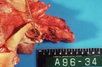 Acute bacterial endocarditis caused by Staphylococcus aureus with perforation of the aortic valve and aortic valve vegetations. Courtesy of Janet Jones, MD, Laboratory Service, Wichita Veterans Administration Medical Center. NextBackground
Infective endocarditis (IE) is defined as an infection of the endocardial surface of the heart, which may include one or more heart valves, the mural endocardium, or a septal defect. Its intracardiac effects include severe valvular insufficiency, which may lead to intractable congestive heart failure and myocardial abscesses. IE also produces a wide variety of systemic signs and symptoms through several mechanisms, including both sterile and infected emboli and various immunological phenomena.[5, 6, 7]
The history of IE can be divided into several eras. In 1674, Lazaire Riviere first described the gross autopsy findings of the disease in his monumental work Opera medica universa. In 1885, William Osler presented the first comprehensive description of endocarditis in English. Lerner and Weinstein presented a thorough discussion of this disease in modern times in their landmark series of articles, “Infective Endocarditis in the Antibiotic Era,†published in 1966 in the New England Journal of Medicine.[8, 9, 10]
IE currently can be described as infective endocarditis in the era of intravascular devices, as infection of intravascular lines has been determined to be the primary risk factor for Staphylococcus aureus bloodstream infections (BSIs). S aureus has become the primary pathogen of endocarditis.[11]
IE generally occurs as a consequence of nonbacterial thrombotic endocarditis, which results from turbulence or trauma to the endothelial surface of the heart. A transient bacteremia then seeds the sterile platelet/fibrin thrombus, with IE as the end result. Pathologic effects due to infection can include local tissue destruction and embolic phenomena. In addition, secondary autoimmune effects, such as immune complex glomerulonephritis and vasculitis, can occur. (See Pathophysiology.)
IE remains a diagnostic and therapeutic challenge. Its manifestations may be muted by the indiscriminate use of antimicrobial agents or by underlying conditions in frail and elderly individuals or immunosuppressed persons. (See Diagnosis.)
Effective therapy has become progressively more difficult to achieve because of the proliferation of implanted biomechanical devices and the rise in the number of resistant organisms. Antibiotic prophylaxis has probably had little effect in decreasing the incidence of IE. (See Treatment and Management.)
For other discussions on IE, see Pediatric Bacterial Endocarditis, Infectious Endocarditis, Neurological Sequelae of Infective Endocarditis, and Antibiotic Prophylactic Regimens for Endocarditis.
Types of infective endocarditisEndocarditis has evolved into several variations, keeping it near the top of the list of diseases that must not be misdiagnosed or overlooked. Endocarditis can be broken down into the following categories:
Native valve endocarditis (NVE), acute and subacuteProsthetic valve endocarditis (PVE),[12] early and late Intravenous drug abuse (IVDA) endocarditisOther terms commonly used to classify types of IE include pacemaker IE and nosocomial IE (NIE).
The classic clinical presentation and clinical course of IE has been characterized as either acute or subacute. Indiscriminate antibiotic usage and an increase in immunosuppressed patients have blurred the distinction between these 2 major types; however, the classification still has clinical merit.[13]
Acute NVE frequently involves normal valves and usually has an aggressive course. It is a rapidly progressive illness in persons who are healthy or debilitated. Virulent organisms, such as S aureus and group B streptococci, are typically the causative agents of this type of endocarditis. Underlying structural valve disease may not be present.
Subacute NVE typically affects only abnormal valves. Its course, even in untreated patients, is usually more indolent than that of the acute form and may extend over many months. Alpha-hemolytic streptococci or enterococci, usually in the setting of underlying structural valve disease, typically are the causative agents of this type of endocarditis.
PVE accounts for 10-20% of cases of IE. Eventually, 5% of mechanical and bioprosthetic valves become infected. Mechanical valves are more likely to be infected within the first 3 months of implantation, and, after 1 year, bioprosthetic valves are more likely to be infected. The valves in the mitral valve position are more susceptible than those in the aortic areas.[12]
Early PVE occurs within 60 days of valve implantation. Traditionally, coagulase-negative staphylococci, gram-negative bacilli, and Candida species have been the common infecting organisms. Late PVE occurs 60 days or more after valve implantation. Staphylococci, alpha-hemolytic streptococci, and enterococci are the common causative organisms. Recent data suggest that S aureus may now be the most common infecting organism in both early and late PVE.[14]
In 75% of cases of IVDA IE, no underlying valvular abnormalities are noted, and 50% of these infections involve the tricuspid valve.[15] S aureus is the most common causative organism.
Analogous to PVE are infections of implantable pacemakers and cardioverter-defibrillators. Usually, these devices are infected within a few months of implantation. Infection of pacemakers includes that of the generator pocket (the most common), infection of the proximal leads, and infection of the portions of the leads in direct contact with the endocardium.
This last category represents true pacemaker IE, is the least common infectious complication of pacemakers (0.5% of implanted pacemakers), and is the most challenging to treat. Of pacemaker infections, 75% are produced by staphylococci, both coagulase-negative and coagulase-positive.
NIE is defined as an infection that manifests 48 hours after the patient is hospitalized or that is associated with a hospital, based on a procedure performed within 4 weeks of clinical disease onset. The term healthcare-associated infective endocarditis (HCIE) is preferable to NIE, since it is inclusive of all sites that deliver patient care, such as hemodialysis centers. The term NIE should be applied to cases of IE acquired in the hospital. An appropriate alternative term would be iatrogenic IE.
Two types of NIE have been described. The right-sided variety affects a valve that has been injured by placement of an intravascular line (eg, Swan-Ganz catheter). Subsequently, the valve is infected by a nosocomial bacteremia. The second type develops in a previously damaged valve and is more likely to occur on the left side. S aureus has been the predominant pathogen of NIE/HCIE since the recent prevalence of intravascular devices. Enterococci are second most commonly isolated pathogens. These usually arise from a genitourinary source.
Evolution of clinical characteristics of infective endocarditisSince the 1960s, the clinical characteristics of IE have changed significantly. The dramatic “graying†of the disease and the increase in recreational drug use and proliferation of invasive vascular procedures underlie this phenomenon. Varieties of IE that were uncommon in the early antibiotic era have become prominent. Cases of NIE, IVDA IE, and PVE have markedly increased. Valvular infections have entered the era of IE caused by intravascular devices and procedures.
The underlying valvular pathology has also changed. Rheumatic heart disease currently accounts for less than 20% of cases, and 6% of patients with rheumatic heart disease eventually develop IE. Approximately 50% of elderly patients have calcific aortic stenosis as the underlying pathology. Congenital heart disease accounts for 15% of cases, with the bicuspid aortic valve being the most common example.
Other contributing congenital abnormalities include ventricular septal defects, patent ductus arteriosus, and tetralogy of Fallot. Atrial septal defect (secundum variety) is rarely associated with IE. Mitral valve prolapse is the most common predisposing condition found in young adults and is the predisposing condition in 30% of cases of NVE in this age group. IE complicates 5% of cases of asymmetrical septal hypertrophy, usually involving the mitral valve.
PreviousNextPathophysiologyIE develops most commonly on the mitral valve, closely followed in descending order of frequency by the aortic valve, the combined mitral and aortic valve, the tricuspid valve, and, rarely, the pulmonic valve. Mechanical prosthetic and bioprosthetic valves exhibit equal rates of infection.
All cases of IE develop from a commonly shared process, as follows:
Bacteremia (nosocomial or spontaneous) that delivers the organisms to the surface of the valveAdherence of the organismsEventual invasion of the valvular leafletsThe common denominator for adherence and invasion is nonbacterial thrombotic endocarditis, a sterile fibrin-platelet vegetation. The development of subacute IE depends on a bacterial inoculum sufficient to allow invasion of the preexistent thrombus. This critical mass is the result of bacterial clumping produced by agglutinating antibodies.
In acute IE, the thrombus may be produced by the invading organism (ie, S aureus) or by valvular trauma from intravenous catheters or pacing wires (ie, NIE/HCIE). S aureus can invade the endothelial cells (endotheliosis) and increase the expression of adhesion molecules and of procoagulant activity on the cellular surface. Nonbacterial thrombotic endocarditis may result from stress, renal failure, malnutrition, systemic lupus erythematosus, or neoplasia.
The Venturi effect also contributes to the development and location of nonbacterial thrombotic endocarditis. This principle explains why bacteria and the fibrin-platelet thrombus are deposited on the sides of the low-pressure sink that lies just beyond a narrowing or stenosis.
In patients with mitral insufficiency, bacteria and the fibrin-platelet thrombus are located on the atrial surface of the valve. In patients with aortic insufficiency, they are located on the ventricular side. In these examples, the atria and ventricles are the low-pressure sinks. In the case of a ventricular septal defect, the low-pressure sink is the right ventricle and the thrombus is found on the right side of the defect.
Nonbacterial thrombotic endocarditis may also form on the endocardium of the right ventricle, opposite the orifice that has been damaged by the jet of blood flowing through the defect (ie, the MacCallum patch).
The microorganisms that most commonly produce endocarditis (ie, S aureus; Streptococcus viridans; group A, C, and G streptococci; enterococci) resist the bactericidal action of complement and possess fibronectin receptors for the surface of the fibrin-platelet thrombus. Among the many other characteristics of IE-producing bacteria demonstrated in vitro and in vivo, some features include the following:
Increased adherence to aortic valve leaflet disks by enterococci, S viridans, and S aureusMucoid-producing strains of S aureusDextran-producing strains of S viridansS viridans and enterococci that possess FimA surface adhesinPlatelet aggregation by S aureus and S viridans and resistance of S aureus to platelet microbicidal proteinsThe pathogenesis of pacemaker IE is similar. Shortly after implantation, the development of a fibrin-platelet thrombus (similar to the nonbacterial thrombotic endocarditis described above) involves the generator box and conducting leads. After 1 week, the connective tissue proliferates, partially embedding the leads in the wall of the vein and endocardium. This layer may offer partial protection against infection during a bacteremia.
Bacteremia (either spontaneous or due to an invasive procedure) infects the sterile fibrin-platelet vegetation described above. BSIs develop from various extracardiac types of infection, such as pneumonias or pyelonephritis, but most commonly from gingival disease. Of those with high-grade gingivitis, 10% have recurrent transient bacteremias (usually streptococcal species). Most cases of subacute disease are secondary to the bacteremias that develop from the activities of daily living (eg, brushing teeth, bowel movements).
The skin is quite resistant to S aureus infection due in great part to its production of antimicrobial peptides. Soong et al discovered that, in vitro, the secretion of alpha toxin by S aureus allows the organism to successfully penetrate the keratinocyte layer. This could explain the presence of staphylococcal bacteremia in the absence of any gross damage to the epithelial layer.[16]
Bacteremia can result from various invasive procedures, ranging from oral surgery to sclerotherapy of esophageal varices to genitourinary surgeries to various abdominal operations. The potential for invasive procedures to produce a bacteremia varies greatly. Procedures, rates, and organisms are as follows:
Endoscopy - Rate of 0-20%; coagulase-negative staphylococci (CoNS), streptococci, diphtheroidsColonoscopy - Rate of 0-20%; Escherichia coli, Bacteroides speciesBarium enema - Rate of 0-20%; enterococci, aerobic and anaerobic gram-negative rodsDental extractions - Rate of 40-100%; S viridansTransurethral resection of the prostate - Rate of 20-40%; coliforms, enterococci, S aureusTransesophageal echocardiography - Rate of 0-20%; S viridans, anaerobic organisms, streptococciThe incidence of nosocomial bacteremias, mostly associated with intravascular lines, has more than doubled in the last few years. Up to 90% of BSIs caused by these devices are secondary to the placement of various types of central venous catheters. Hickman and Broviac catheters are associated with the lowest rates, presumably because of their Dacron cuffs. Peripherally placed central venous catheters are associated with similar rates.
Intravascular catheters are infected from 1 of the following 4 sources:
Infection of the insertion siteInfection of the catheterBacteremia arising from another siteContamination of the infused solutionBacterial adherence to intravascular catheters depends on the response of the host to the presence of this foreign body, the properties of the organism itself, and the position of the catheter. Within a few days of insertion, a sleeve of fibrin and fibronectin is deposited on the catheter. S aureus adheres to the fibrin component.
S aureus also produces an infection of the endothelial cells (endotheliosis), which is important in producing the continuous bacteremia of S aureus BSIs. Endotheliosis may explain many cases of persistent methicillin-susceptible S aureus (MSSA) and methicillin-resistant S aureus (MRSA) catheter-related BSIs without an identifiable cause.
S aureus catheter-related BSIs occur even after an infected catheter is removed, apparently attributable to specific virulence factors of certain strains of S aureus that invade the adjacent endothelial cells. At some point, the staphylococci re-enter the bloodstream, resulting in bacteremia.[17]
Four days after placement, the risk of infection markedly increases. Lines positioned in the internal jugular are more prone to infection than those placed in the subclavian vein. Colonization of the intracutaneous tract is the most likely source of short-term catheter-related BSIs. Among lines in place for more than 2 weeks, infection of the hub is the major source of bacteremia. In some cases, the infusion itself may be a reservoir of infection.
Colonization of heart valves by microorganisms is a complex process. Most transient bacteremias are short-lived, are without consequence, and are often not preventable. Bacteria rarely adhere to an endocardial nidus before the microorganisms are removed from the circulation by various host defenses.
Once microorganisms do establish themselves on the surface of the vegetation, the process of platelet aggregation and fibrin deposition accelerate at the site. As the bacteria multiply, they are covered by ever-thickening layers of platelets and thrombin, which protect them from neutrophils and other host defenses. Organisms deep in the vegetation hibernate because of the paucity of available nutrients and are therefore less susceptible to bactericidal antimicrobials that interfere with bacterial cell wall synthesis.
Complications of subacute endocarditis result from embolization, slowly progressive valvular destruction, and various immunological mechanisms. The pathological picture of subacute IE is marked by valvular vegetations in which bacteria colonies are present both on and below the surface.
The cellular reaction in SBE is primarily that of mononuclear cells and lymphocytes, with few polymorphonuclear cells. The surface of the valve beneath the vegetation shows few organisms. Proliferation of capillaries and fibroblasts is marked. Areas of healing are scattered among areas of destruction. Over time, the healing process falls behind, and valvular insufficiency develops secondary to perforation of the cusps and damage to the chordae tendineae. Compared with acute disease, little extension of the infectious process occurs beyond the valvular leaflets.
levels of agglutinating and complement-fixing bactericidal antibodies and cryoglobulins are markedly increased in patients with subacute endocarditis. Many of the extracardiac manifestations of this form of the disease are due to circulating immune complexes. Among these include glomerulonephritis, peripheral manifestations (eg, Osler nodes, Roth spots, subungual hemorrhages), and, possibly, various musculoskeletal abnormalities. Janeway lesions usually arise from infected microemboli.
The microscopic appearance of acute bacterial endocarditis differs markedly from that of subacute disease. Vegetations that contain no fibroblasts develop rapidly, with no evidence of repair. Large amounts of both polymorphonuclear leukocytes and organisms are present in an ever-expanding area of necrosis. This process rapidly produces spontaneous rupture of the leaflets, of the papillary muscles, and of the chordae tendineae.
The complications of acute bacterial endocarditis result from intracardiac disease and metastatic infection produced by suppurative emboli. Because of their shortened course, immunological phenomena are not a part of acute IE.
PreviousNextEtiologyThe different types of IE have varying causes and involve different pathogens.
Native valve endocarditisThe following are the main underlying causes of NVE:
Rheumatic valvular disease (30% of NVE) - Primarily involves the mitral valve followed by the aortic valveCongenital heart disease (15% of NVE) - Underlying etiologies include a patent ductus arteriosus, ventricular septal defect, tetralogy of Fallot, or any native or surgical high-flow lesion. Mitral valve prolapse with an associated murmur (20% of NVE)Degenerative heart disease - Including calcific aortic stenosis due to a bicuspid valve, Marfan syndrome, or syphilitic diseaseApproximately 70% of infections in NVE are caused by Streptococcus species, including S viridans, Streptococcus bovis, and enterococci. Staphylococcus species cause 25% of cases and generally demonstrate a more aggressive acute course (see the images below).
Prosthetic valve endocarditisEarly PVE, which presents shortly after surgery, has a different bacteriology and prognosis than late PVE, which presents in a subacute fashion similar to NVE.
Infection associated with aortic valve prostheses is particularly associated with local abscess and fistula formation, and valvular dehiscence. This may lead to shock, heart failure, heart block, shunting of blood to the right atrium, pericardial tamponade, and peripheral emboli to the central nervous system and elsewhere.
Early PVE may be caused by a variety of pathogens, including S aureus and S epidermidis. These nosocomially acquired organisms are often methicillin-resistant (eg, MRSA).[18] Late disease is most commonly caused by streptococci. Overall, CoNS are the most frequent cause of PVE (30%).
S aureus causes 17% of early PVE and 12% of late PVE. Corynebacterium, nonenterococcal streptococci, fungi (eg, C albicans, Candida stellatoidea, Aspergillus species), Legionella, and the HACEK (ie, Haemophilus aphrophilus, Actinobacillus actinomycetemcomitans, Cardiobacterium hominis, Eikenella corrodens, Kingella kingae) organisms cause the remaining cases.
IVDA infective endocarditisDiagnosis of endocarditis in IV drug users can be difficult and requires a high index of suspicion. Two thirds of patients have no previous history of heart disease or murmur on admission. A murmur may be absent in those with tricuspid disease, owing to the relatively small pressure gradient across this valve. Pulmonary manifestations may be prominent in patients with tricuspid infection: one third have pleuritic chest pain, and three quarters demonstrate chest radiographic abnormalities.
S aureus is the most common (S aureus infections and has been associated with previous hospitalizations, long-term addiction, and nonprescribed antibiotic use. Groups A, C, and G streptococci and enterococci are also recovered from patients with IVDA IE.
Currently, gram-negative organisms are involved infrequently. P aeruginosa[19] and the HACEK family are the most common examples.
Nosocomial/healthcare-associated infective endocarditisEndocarditis may be associated with new therapeutic modalities involving intravascular devices such as central or peripheral intravenous catheters, rhythm control devices such as pacemakers and defibrillators, hemodialysis shunts and catheters, and chemotherapeutic and hyperalimentation lines.[20, 21] These patients tend to have significant comorbidities, more advanced age, and predominant infection with S aureus. The mortality rate is high in this group.
The organisms that cause NIE/HCIE obviously are related to the type of underlying bacteremia. The gram-positive cocci (ie, S aureus, CoNS, enterococci, nonenterococcal streptococci) are the most common pathogens.
Fungal endocarditisFungal endocarditis is found in intravenous drug users and intensive care unit patients who receive broad-spectrum antibiotics.[22] Blood cultures are often negative, and diagnosis frequently is made after microscopic examination of large emboli.
Clinical features associated with different pathogensDifferent causative organisms tend to give rise to varying clinical manifestations of IE, as shown in the Table below.
Table 1. Clinical Features of Infective Endocarditis According to Causative Organism (Open Table in a new window)
Causative Organism(s) Clinical Features of IE Staphylococcus aureusOverall, S aureus infection is the most common cause of IE, including PVE, acute IE, and IVDA IE.Approximately 35-60.5% of staphylococcal bacteremias are complicated by IE.More than half the cases are not associated with underlying valvular disease.The mortality rate of S aureus IE is 40-50%.S aureus infection is the second most common cause of nosocomial BSIs, second only to CoNS infection.The incidence of MRSA infections, both the hospital- and community-acquired varieties, has dramatically increased (50% of isolates). Sixty percent of individuals are intermittent carriers of MRSA or MSSA .The primary risk factor for S aureus BSI is the presence of intravascular lines. Other risk factors include cancer, diabetes, corticosteroid use, IVDA, alcoholism, and renal failure. The realization that approximately 50% of hospital- and community-acquired staphylococcal bacteremias arise from infected vascular catheters has led to the reclassification of staphylococcal BSIs. BSIs are acquired not only in the hospital but also in any type of health care facility (eg, nursing home, dialysis center). Of S aureus bacteremia cases in the United States, 7.8% (200,000) per year are associated with intravascular catheters.Streptococcus viridansThis organism accounts for approximately 50-60% of cases of subacute disease.Most clinical signs and symptoms are mediated immunologically.Streptococcus intermedius groupThese infections may be acute or subacute.S intermedius infection accounts for 15% of streptococcal IE cases.Members of the S intermedius group, especially S anginosus, are unique among the streptococci in that they can actively invade tissue and form abscesses, often in the CNS.AbiotrophiaApproximately 5% of subacute cases of IE are due to infection with Abiotrophia species.They require metabolically active forms of vitamin B-6 for growth.This type of IE is associated with large vegetations that lead to embolization and a high rate of posttreatment relapse.Group D streptococciMost cases are subacute.The source is the gastrointestinal or genitourinary tract.It is the third most common cause of IE.They pose major resistance problems for antibiotics.Nonenterococcal group DThe clinical course is subacute.Infection often reflects underlying abnormalities of the large bowel (eg, ulcerative colitis, polyps, cancer).The organisms are sensitive to penicillin.Group B streptococciAcute disease develops in pregnant patients and older patients with underlying diseases (eg, cancer, diabetes, alcoholism).The mortality rate is 40%.Complications include metastatic infection, arterial thrombi, and congestive heart failure.It often requires valve replacement for cure.Group A, C, and G streptococciAcute disease resembles that of S aureus IE (30-70% mortality rate), with suppurative complications.Group A organisms respond to penicillin alone.Group C and G organisms require a combination of synergistic antibiotics (as with enterococci).Coagulase-negative S aureusThis causes subacute disease.It behaves similarly to S viridans infection.It accounts for approximately 30% of PVE cases and less than 5% of NVE cases.[23] Pseudomonas aeruginosaThis is usually acute, except when it involves the right side of the heart in IVDA IE.Surgery is commonly required for cure.HACEK (ie, Haemophilus aphrophilus, Actinobacillus actinomycetemcomitans, Cardiobacterium hominis, Eikenella corrodens, Kingella kingae)These organisms usually cause subacute disease.They account for approximately 5% of IE cases.They are the most common gram-negative organisms isolated from patients with IE.Complications may include massive arterial emboli and congestive heart failure.Cure requires ampicillin, gentamicin, and surgery.FungalThese usually cause subacute disease.The most common organism of both fungal NVE and fungal PVE is Candida albicans.Fungal IVDA IE is usually caused by Candida parapsilosis or Candida tropicalis.Aspergillus species are observed in fungal PVE and NIE.BartonellaThe most commonly involved species is Bartonella quintana.IE typically develops in homeless males who have extremely substandard hygiene. Bartonella must be considered in cases of culture-negative endocarditis among homeless individuals.Multiple pathogens (polymicrobial)Pseudomonas and enterococci are the most common combination of organisms.It is observed in cases of IVDA IEThe cardiac surgery mortality rate is twice that associated with single-agent IE.[24] Risk factorsThe most significant risk factor for IE is residual valvular damage caused by a previous attack of endocarditis.[25, 20]
Many possible risk factors for the development of pacemaker IE have been described, including diabetes mellitus, age, and use of anticoagulants and corticosteroids. The evidence for these is conflicting. The major risk factor is probably surgical intervention to any part of the pacemaker system, especially elective battery replacements. The rate of infection associated with battery replacements is approximately 5 times that of the initial implantation (6.5% vs 1.4%).
Other significant risk factors for pacemaker IE include the development of a postoperative hematoma, the inexperience of the surgeon, and a preceding temporary transvenous pacing.
PreviousNextEpidemiologyIn the United States, the 2009 incidence of IE was approximately 12.7 cases per 100,000 persons per year.[26] The age-adjusted hospital admission rate has increased 2.4% annually from 1998-2009. This rate has risen significantly from that of the previous 50 years (2-4 cases per 100,000 persons per year).[27] The incidence of IE in other countries is similar to that in the United States. From 1998-2009, the proportion of patients with intracardiac devices increased from 13.3% to 18.9%, while the proportion of cases with a background of HIV infection or HIV drug abuse fell.
Between 1998 and 2009, the mean age of patients has risen from 58.6 to 60.8 years.[26] Currently, more than 50% of patients are older than 50 years.[20] Mendiratta et al, in their retrospective study of hospital discharges from 1993-2003 of patients aged 65 years and older with a primary or secondary diagnosis of IE, found that hospitalizations for IE increased 26%, from 3.19 per 10,000 elderly patients in 1993 to 3.95 per 10,000 in 2003.[28] This increase in age has continued, with the mean age of patients in 2009 at 60.8 years.[26]
IE is 3 times as common in males as in females. It has no racial predilection.
PreviousNextPrognosisPrognosis largely depends on whether or not complications develop. If left untreated, IE is generally fatal. Early detection and appropriate treatment of this uncommon disease can be lifesaving. The overall mortality rate has remained stable at 14.5%.[26]
Cure rates for appropriately managed (including both medical and surgical therapies) NVE are as follows:
For S viridans and S bovis infection, the rate is 98%.For enterococci and S aureus infection in individuals who abuse intravenous drugs, the rate is 90%.For community-acquired S aureus infection in individuals who do not abuse intravenous drugs, the rate is 60-70%.For infection with aerobic gram-negative organisms, the rate is 40-60%.For infection with fungal organisms, the rate is lower than 50%.For PVE, the cure rates are as follows:
Rates are 10-15% lower for each of the above categories, for both early and late PVE.Surgery is required far more frequently.Approximately 60% of early CoNS PVE cases and 70% of late CoNS PVE cases are curable.Anecdotal reports describe the resolution of right-sided valvular infection caused by S aureus infection in individuals who abuse intravenous drugs after just a few days of oral antibiotics.
The role of early valvular surgery in reducing mortality among patients with IE has become somewhat clearer. Challenges to resolving this question include the necessity of performing multicentered studies with an apparent difficulty of ensuring that the patients' preoperative assessments and surgical approaches are comparable. The largest study to date indicates that in cases of IE complicated by heart failure, valvular surgery reduces the 1-year mortality rate.[29] More recent studies document that early surgery in patients, especially those with large vegetations, significantly reduces the risk of death from any cause that from embolic events.[30, 31]
Mortality rates in NVE range from 16-27%. Mortality rates in patients with PVE are higher. More than 50% of these infections occur within 2 months after surgery. The fatality rate of pacemaker IE ranges up to 34%.[32]
Increased mortality rates are associated with increased age,[33] infection involving the aortic valve, development of congestive heart failure, central nervous system (CNS) complications, and underlying disease such as diabetes mellitus. Catastrophic neurological events of all types due to IE are highly predictive of morbidity and mortality.[34]
Mortality rates also vary with the infecting organism. Acute endocarditis due to S aureus is associated with a high mortality rate (30-40%), except when it is associated with IV drug use.[14, 35] Endocarditis due to streptococci has a mortality rate of approximately 10%.
PreviousNextPatient EducationSurveys indicate that an appallingly small number of patients who are at risk for developing IE have an understanding of antibiotic and nonpharmacologic (ie, appropriate oral hygiene) principles. Drug rehabilitation for patients who use IV drugs is critical.
The United Kingdom’s National Institute for Health and Clinical Excellence (NICE) addresses patient education in its 2008 guideline on prophylaxis against IE in adults and children undergoing interventional procedures. The NICE’s guideline recommends that health care professionals teach patients about the symptoms of IE and the risks of nonmedical invasive procedures such as body piercing and tattooing, explain the benefits and risks of antibiotic prophylaxis and the reasons that it is no longer routine, and emphasize the need to maintain good oral health.[36]
For patient education information, see the Heart Center, as well as Tetralogy of Fallot.
PreviousProceed to Clinical Presentation , Infective Endocarditis


0 comments:
Post a Comment